2238
Different patterns of cortical matter changes in first episode bipolar manic adolescents: a surface-based structural MRI study with cluster analysis1Huaxi MR Research Center (HMRRC), Department of Radiology, West China Hospital of Sichuan University, Chengdu, People's Republic of China, 2Department of Psychiatry and Behavioral Neuroscience, University of Cincinnati College of Medicine, Cincinnati, OH, United States
Synopsis
With an objective neuroimaging data-driven method using agglomerative hierarchical clustering analysis, two patterns of cortical matter changes among 58 first episode bipolar manic adolescents has been identified. And the clinical feature especially the intelligence quotient was corresponding to the subtyping among these patients. While the effect of medication and illness duration have been minimized, our findings provided new evidence indicating the existence of two neurobiologically distinct subgroups of patients with first episode bipolar mania, which may reflect qualitatively distinct genetic influences or neurodevelopmental alterations.
INTRODUCTION
Structural gray matter abnormalities are crucial to the neurobiological models of the bipolar disorder by providing quantitative measures of brain tissue, however, findings from the existing neuroimaging studies are quite inconsistent. Besides the confounders including illness duration, medication effects and comorbidities, the heterogeneity of the disorder in pathophysiology contributed greatly to the discrepancy and limited the clinical translations of these imaging findings [1]. By adopting an objective data-driven method, the present study investigated whether different patterns of cortical matter changes exist in a group of first episode bipolar manic adolescents.METHODS
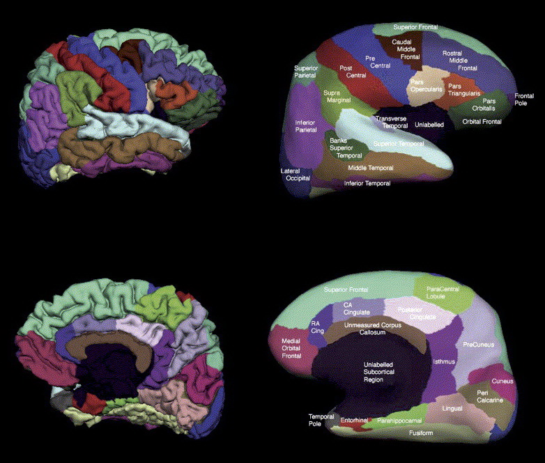
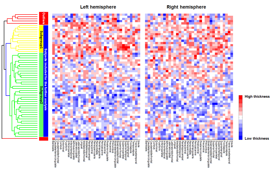
he study was approved by the local research ethics committee, and all study participants provided written assent with written informed consent from their legal guardians as they all were less than 18 years at age. In a cross-sectional design, high-resolution T1-weighted images were acquired from 58 first episode manic adolescents and 31 well-matched healthy controls. Bipolar subjects were regarded as first-episode if they were currently experiencing or had recently recovered from their first manic or mixed episode as defined in DSM-IV-TR criteria. Psychopathological ratings were assessed using Young Mania Rating Scale (YMRS) and Children’s Depression Rating Scale (CDRS). Age, sex and education level were well matched between diagnostic groups. MRI examinations were performed on a 4-T Varian Unity INOVA scanner with Modified Driven Equilibrium Fourier Transform (MDEFT) pulse sequence (TR=13.0 ms, TE=5.3 ms, tau (magnetization preparation time) =1.1 ms, FOV=256×192×192 mm3, matrix=256×192×96). FreeSurfer (version 5.3.0, http://surfer.nmr.mgh.harvard.edu/) was adopted for the constructions of cortical surface and volumetric segmentation, which were developed from three dimensional spoiled gradient recalled images. Firstly, agglomerative hierarchical clustering [2] was performed on bipolar manic patients to detect whether there are subgroups among these patients, and cortical thickness of all the regions within Desikan Atlas [3] in bilateral hemispheres were calculated and selected as the features for the cluster analysis across patients. Secondly, point-wise cortical comparisons were conducted among the potential patient subgroups identified with cluster analysis, if there were, and healthy comparisons.RESULTS
With cortical morphometric features extracted from 68 main cortical regions (Figure 1), two main subgroups of patients were identified, while the other 6 subjects were regarded as outliers (Figure 2). We tried the analysis without the outliers and the findings were replicated. Besides, patients within subgroup 1 showed significant differences in intelligence quotient (IQ) and social economic status relative to control subjects, whilst those of subgroup 2 did not. Subjects in subgroup 1 showed widespread cortical thickening when compared with health controls, mainly in fronto-temporo-parietal regions, whereas subgroup 2 only showed regional cortical thinning in left superior temporal gyrus and left superior parietal cortex. The cortical thickness of left caudal middle frontal gyrus is negative associated with YMRS total scores, whilst that of right pars orbitalis is negative associated with CDRS total scores.DISCUSSION
By combining quantitative measures and an objective neuroimaging data-driven analysis on a group of purified first episode bipolar manic patients, two distinct patterns of cortical matter abnormality were identified among those subjects. One of the subgroups showed widespread cortical thickening in fronto-temporo-parietal regions, which might suggest potential neuronal disorganization in these regions. As synaptic pruning is a process which would sculpt the brain into maturity and expanded its capacity for high-level reasoning, disturbance in this process could occur as a consequence of a defective maturational shaping. That is overlapped with the observation that patients in subgroup 1 have lower IQ than control subjects, while patients in subgroup 2 does not. The subgroup 2 showed more localized cortical thinning in the left superior temporal gyrus and left superior parietal cortex, regions have been frequently observed in this disorder and may appear to be a familial trait, which begin at the prodromal states or before illness onset [5].CONCLUSION
Two distinct patterns of cortical matter abnormalities were identified, suggesting the existence of different pathophysiologic processes related to brain morphometric changes. While the effect of medication and illness duration have been minimized, our findings provided new evidence indicating the existence of two neurobiologically distinct subgroups of patients with first episode bipolar mania, which may reflect qualitatively distinct genetic influences or neurodevelopmental alterations. Future probing with data of other modalities including cognitive, neuropathological and genetic information may optimize the classification and give a better insight into the specific pathophysiological substrate of each subgroup.Acknowledgements
This work was supported by National Natural Science Foundation of China (Grant numbers 81371527, 81671664, 81621003) and National Program for Support of Top-notch Young Professionals.References
1. Hozer F, Houenou J. Can neuroimaging disentangle bipolar disorder? Journal of affective disorders. 2016;195:199-214.
2. Johnson SC. Hierarchical clustering schemes. Psychometrika. 1967;32(3):241-254.
3. Salat DH, Buckner RL, Snyder AZ et al. Thinning of the cerebral cortex in aging. Cereb Cortex. 2004;14:721-730.
4. Narr KL, Toga AW, Szeszko P et al. Cortical thinning in cingulate and occipital cortices in first episode schizophrenia. Biol Psychiatry. 2005;58:32-40.
5. Hanford LC, Nazarov A, Hall GB et al. Cortical thickness in bipolar disorder: a systematic review. Bipolar disorders. 2016;18:4-18.
Figures