2232
Cerebral blood flow measurements in patients with depression and comorbid hypertension in depression using 3D arterial spin labeling1Radiology, Peking University Third Hospital, Beijing, People's Republic of China, 2GE Healthcare, MR Research China, Beijing, People's Republic of China
Synopsis
The aim of this study is to explore the potential differences of the quantitative CBF values among patients with depression, patients with hypertension and patients with comorbid hypertension in depression by using whole brain 3D PCASL and to investigate the correlation between CBF values and degrees of depression. The results indicate that CBF values decrease in frontal and parietal lobes in patients with comorbid hypertension in depression, whereas not in patients with depression or patients with hypertension, hypertension may play a synergistic action in the progress of depression.
Purpose:
Depressive disorder is a mental disease with typical clinical manifestations such as depressed mood and mental retardation. Decreases of cerebral blood flow (CBF) and cerebral glucose metabolism have been observed in certain cerebral areas such as the cingulated, frontal, temporal and parietal regions [1]. The incidence of hypertension increases with age, hence, depression patients that are observed with comorbid hypertension are diagnosed as hypertension comorbid in depression. Arterial spin labeling (ASL) is a noninvasive MR technique that can be utilized for the measurement of CBF, which employs magnetically labeled arterial blood water as the endogenous contrast medium [2]. However, there has not been any article reporting CBF perfusion in patients with comorbid hypertension in depression or the influence of hypertension on depression by using three dimensional(3D) pseudo-continuous arterial spin labeling (PCASL). The aim of this study was to explore the potential differences of the quantitative CBF values among patients with depression, patients with hypertension and patients with comorbid hypertension in depression by using whole brain 3D PCASL and to investigate the potential correlation between CBF values and the degrees of depression.Material and Method:
The current study recruited sixteen patients with depression (2 males and 14 females, age range 42~72 years old, mean age 58.1±8.1 years old), twenty patients with hypertension (4 males and 16 females, age range 41~65 years old, mean age 58.0±5.4 years old), sixteen patients with comorbid hypertension in depression (3males and 13 females, age range 45~74 years old, mean age 61.0±6.4 years old) and twenty healthy control subjects(HC) (4males and 16 females, age range 43~68 years old, mean age 56.1±8.6 years old).3D PCASL was performed on a 3.0T MR scanner(Discovery750, GE Healthcare, Milwaukee, USA) with an eight-channel head coil applied. Structural images were achieved for anatomic information using a sagittal 3D T1 SPGR sequence, with the following parameters: TR=4.9ms, TE=2ms, FOV=24×24cm, matrix=240×240, 15°flip angle, voxel size=1×1×1mm3, and acquisition time of 6min34sec. 3D PCASL sequence was acquired with parameters as follows: TR=4632ms, TE=10.5ms, FOV=24×24cm, voxel size=2×2×4mm3, post label delay (PLD) =1.5s, and acquisition time of 3min15sec. Statistical parametric mapping (SPM8) was performed to preprocess CBF map. Comparisons of CBF values among four groups were conducted, and the potential correlation between CBF and degrees of depression were investigated.Results:
1. Compared with HC, decreases of CBF values in bilateral middle frontal gyrus were observed in patients with depression, but without significant statistical differences. Compared with HC, there was no significant statistical difference observed in patients with hypertension.
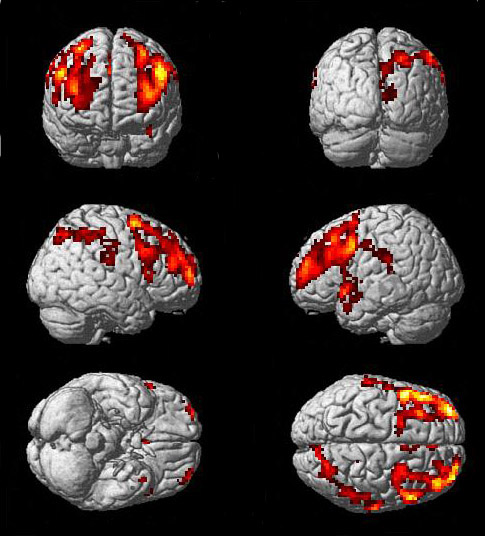
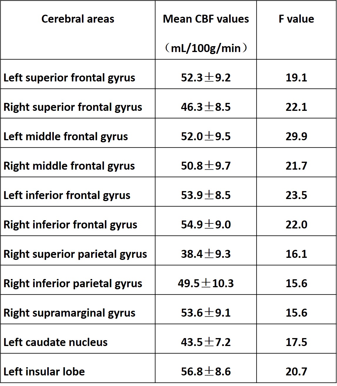
2. On the other hand, patients with comorbid hypertension in depression demonstrated relatively decreased CBF values in bilateral superior frontal gyri, bilateral middle frontal gyri, bilateral inferior frontal gyri, right superior parietal gyrus, right inferior parietal gyrus, right supramarginal gyrus, left caudate nucleus and left insular lobe(P=0.001,F=11.81) as compared with HC ( Figure A and Table1)
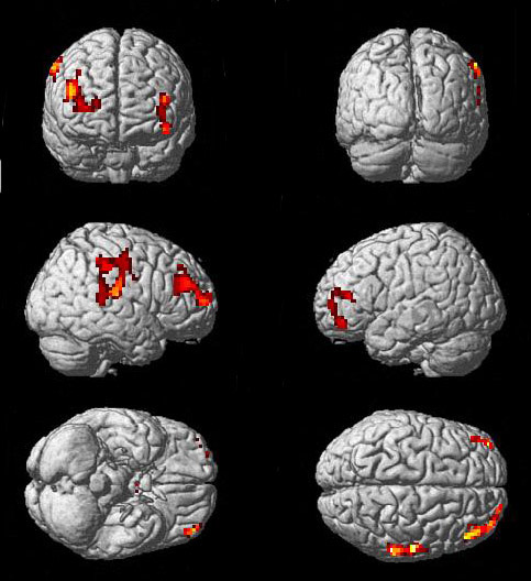
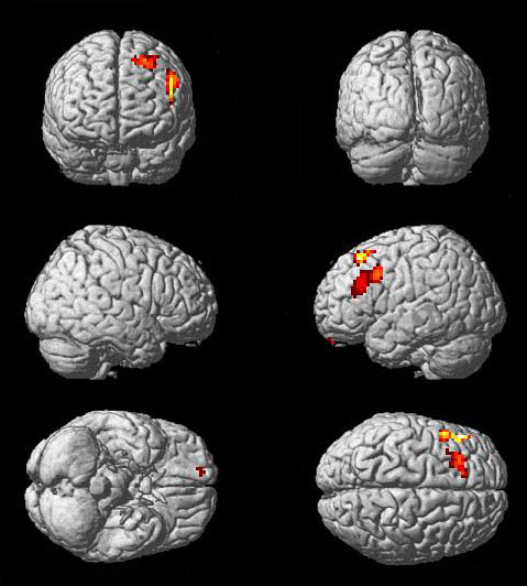
3. Compared with patients with depression, patients with comorbid hypertension in depression exhibited decreased CBF values in right middle frontal gyrus, right supramarginal gyrus and left superior frontal gyrus (P=0.001,F=11.81)( Figure B). Moreover, while compared with patients with hypertension, patients with comorbid hypertension in depression showed lower CBF values in left middle frontal gyrus(P=0.001,F=11.81)( Figure C).
4. However, there was no correlation detected between the decreased CBF values and HAMD scores in any of the four groups (P>0.05).
Conclusions:
Decreases in CBF values were detected in frontal and parietal lobes in patients with comorbid hypertension in depression by using 3D PCASL, whereas not in patients with depression or patients with hypertension. Hypertension may play a synergistic action in promoting the progress of depression. Therefore, it may be essential to consider and to eliminate the effect of blood pressure in patients for the future study of depression.Acknowledgements
No acknowledgement found.References
[1] Colloby SJ, Firbank MJ, He J, et al. Regional cerebral blood flow in late-life depression: arterial spin labelling magneticresonance study. Br J Psychiatry, 2012, 200(2):150-155.
[2] Nezamzadeh M, Matson GB, Young K, et al. Improved pseudo-continuous arterial spin labeling for mapping brain perfusion. J Magn Reson Imaging, 2010, 31(6):1419-1427.
Figures