2223
Imaging Characteristics According to the IDH Mutation and 1p19q Codeletion Status of Lower Grade Gliomas1Yonsei University College of Medicine, Seoul, Korea, Republic of
Synopsis
Preoperative prediction of genotypic classification with MRI is useful because IDH mutation and 1p/19q codeletion status are important prognostic and predictive factors in lower-grade gliomas (WHO grade II and III). In this study we found that various preoperative imaging features including lobar/nonlobar or central location, enhancement characteristics, tumor margin, diffusion characteristics, ependymal/cortical involvement were different between IDH-wildtype and IDH-mutant group. Therefore preoperative MRI may be helpful to predict IDH mutation status in lower-grade gliomas.
Purpose
It has been known that IDH mutation and 1p/19q codeletion status in addition to histology are important prognostic and predictive factors in lower-grade gliomas (WHO grade II and III).1 The purpose of this study was to evaluate the predictive value of imaging features assessed with Visually AcceSAble Rembrandt Images (VASARI) 2 lexicon in lower-grade gliomas according to genotypic classification.Materials and Methods
The preoperative MRI of 179 lower-grade glioma patients with known IDH mutation and 1p/19q codeletion status were included in this study. Among them, 87 patients were WHO grade II and 92 patients were grade III gliomas. According to molecular categories, 77 patients were IDH-wildtype, 55 patients were IDH-mutant and no 1p/19q codeletion, 47 patients were IDH-mutant and 1p/19q codeleted. Preoperative MRI was performed at 3.0 T MRI (Achieva, Philips Medical Systems, Netherlands) with an eight-channel sensitivity-encoding head coil. The preoperative MR imaging protocol included T1-weighted (repetition time msec/echo time msec, 2000/10; field of view, 240 mm; section thickness, 5 mm; matrix, 256 × 256), T2-weighted (3000/80; field of view, 240 mm; section thickness, 5 mm; matrix, 256 × 256), and fluid-attenuated inversion recovery (10 000/125; field of view, 240 mm; section thickness, 5 mm; matrix, 256 × 256) images. Three-dimensional contrast material–enhanced T1-weighted images (6.3/3.1; field of view, 240 mm; section thickness, 1 mm; matrix, 192 × 192) were acquired after administering 0.1 mL/kg gadolinium-based contrast material (Gadovist; Bayer, Toronto, Ontario, Canada). Diffusion-tensor imaging was performed with b values of 600 sec/mm2 and 0 sec/mm2, 32 directions, and the following parameters: 8413.4/77; field of view, 220 mm; section thickness, 2 mm; and matrix, 112 × 112. Two neuroradiologists who were blinded to the molecular data reviewed the conventional MRI for tumor size, location, and tumor morphology by using a standardized imaging feature set VASARI. Imaging features were correlated with IDH mutation status using chi-square statisticial analysis. In addition, imaging features of 1p/19q codeleted group and no 1p/19q codeletion group were compared in the IDH-mutant subgroup.Results
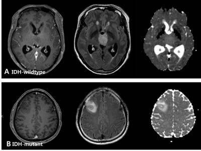
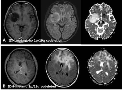
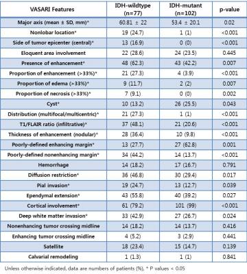
Various preoperative imaging features were different between IDH-wildtype and IDH-mutant group. IDH-wildtype gliomas were larger (major axis) and more frequently showed nonlobar and central location, larger proportion of enhancement/edema/necrosis, multifocal or multicentric distribution, T1/FLAIR ratio (infiltrative), nodular enhancement, poorly-defined nonenhancing margin, diffusion restriction, pial invasion, ependymal extension, and deep white matter invasion (Table) (Figure 1). On the other hand, intratumoral cyst, poorly-defined enhancing margin, and cortical involvement were more common in IDH-mutant gliomas. In the IDH-mutant subgroup, only diffusion characteristic was different between no 1p19q codeletion group and 1p19q codeleted group. IDH-mutant, no 1p19q codeletion group showed predominantly facilitated diffusion characteristics compared with the IDH-mutant, 1p19q codeleted group (Figure 2).Discussion
Our data show that various MRI features of VASARI, including lobar/nonlobar or central location, enhancement characteristics, tumor margin, diffusion characteristics, ependymal/cortical involvement can assist prediction of genotypic classification of lower-grade gliomas preoperatively. Preoperative prediction of genotypic classification of lower-grade glioma with MRI is useful because the overall survival is different according to IDH mutation and chemosensitivity is different according to 1p/19q codeletion status.3Conclusion
MRI features are different according to the molecular markers in lower-grade glioma and may be helpful to predict IDH mutation status preoperatively.Acknowledgements
No acknowledgement found.References
1. Cancer Genome Atlas Research Network. "Comprehensive, integrative genomic analysis of diffuse lower-grade gliomas." N Engl J Med 2015.372 (2015): 2481-2498.
2. https://wiki.nci.nih.gov/display/CIP/VASARI
3. Kaloshi, G., et al. "Temozolomide for low-grade gliomas Predictive impact of 1p/19q loss on response and outcome." Neurology 68.21 (2007): 1831-1836.
Figures


