2218
Application of High B-value Diffusion-Weighted Imaging in Defining High-grade Glioma Infiltration Regions1MRI Room,Department of Imaging Center, The First Affiliated Hospital of Xinjiang Medical University, Urumqi, People's Republic of China, 2MRI Room,Department of Imaging Center, The First Affiliated Hospital of Xinjiang Medical University, 3Department of Neurosurgery, The First Affiliated Hospital of Xinjiang Medical University
Synopsis
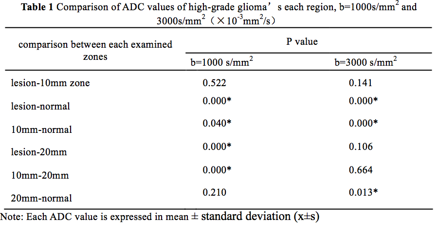
Objective To evaluate the role of the ADC value in patients with high-grade glioma (WHO III-IV) using high B-value diffusion weighted imaging (DWI), as a potential noninvasive quantitative index in defining the boundary of glioma. Methods 25 cases of surgical pathologically confirmed glioma underwent the DWI scan with b=1000 and 3000s/mm2 respectively in a 3T MR scanner. The ADC values of the glioma substantial zone, cerebral parenchyma within 0-10/10-20mm radium and the cerebral parenchyma of the opposite sphere are statistically analyzed by SPSS17.0. Results When b=1000 and 3000 s/mm2, the difference in the ADC values of the tumor and the 0-10mm region had no statistical significance(P>0.05). When b-value = 1000 s/mm2, there is statistically valid difference among ADC values of 10-20mm area, lesions and comparing group (P<0.05); When b-value =3000 s/mm2, there is no statistically valid difference among ADC values of 10-20mm area and lesions(P>0.05). The ADC value of 10-20mm area is validly lower than the lesions. Conclusion The ADC value in high B value DWI scan indicating that the ADC value is more sensitive in quantitatively analyzing the infiltration zone of glioma relative to the low B value DWI scan.
Target audience
Researchers and clinicians interested in brain imaging
and diseases, with a particular interest in diffusion imaging.Introduction
Glioma is one of the most common primary intracranial tumors. Surgical removal is the most effective treatment available at the moment. However, glioma is infiltrative and is very difficult to be completely removed through surgery. It can easily recur and would severely affect post-treatment recovery. Currently the diagnosis and differential diagnosis of glioma relies mainly on imaging results. Studies have shown that traditional MRI can overestimate or underestimate tumor boundaries by 40%[1] . As an improved MRI technique, diffusion-weighted imaging (DWI) is considered the most sensitive for early pathological changes. The sensitivity of DWI to water molecular infiltration increases with its b value. The DWI with higher b value is able to provide better contrast. High b-value DWI is becoming more widely used in such areas as the grading[2-4] and distinguishing[5] of glioma. However, reports concerning the definition of the scope of glioma infiltration are seldom mentioned. Therefore, this study aims to use the strong 3.0T MRI to carry out the DWI b=1000 s/mm2 and b=3000 s/mm2 DWI, and combine them with regular MRI scan and enhanced scan in order to provide a basis decision making solution for defining the boundaries of glioma infiltration.Method
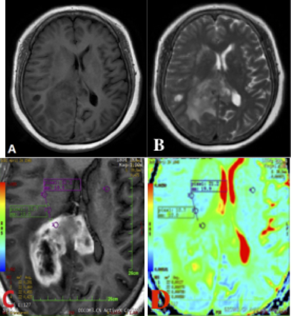
This prospective study was approved by the Ethics Committee and informed consents were obtained from all participants. Twenty-five cases of surgical pathologically confirmed high-grade glioma(WHO III-IV) underwent the DWI scan with b=1000 and 3000s/mm2 respectively in a 3.0T MR scanner (Signa HDXT, GE Healthcare, Milwaukee, USA ). DWI was conducted twice. The scan position was the s-axis, using the Spin-Echo-Echo Planar Imaging(SE-EPI). The slice thickness is 5.0cm, and the slice gap 0.0cm. The number of slices is 26, FOV is 24cm, NEX is 2, the matrix is 128*128, TR is 6000ms, TE is 60ms, and the value of gradient sensitive factor b is 1000s/mm2 or 3000s/mm2. Diffusion gradients will be disbursed over phase encoding, slice selection and output direction (frequency code direction). All workstation images were post processed through GE Function Tool 6.3 via GE ADW4.4 in order to examine the DWI ADC value when b=1000s/mm2 and 3000s/mm2, respectively. With radiologists, we first scanned the x-axis of the slice with the greatest tumor radium, and then placed the Region of Interest (ROI) in the substantial area of the enhanced tumor, in the substantial area within the 0-10mm region outside the boundary of the enhanced tumor, in the substantial are within the 10-20mm region outside the boundary of the enhanced tumor as well as in the substantial area of the corresponding normal region in the opposite sphere.Results
When b=1000 and 3000 s/mm2, the difference in the ADC values of the tumor and the 0-10mm region had no statistical significance(P>0.05). When b-value = 1000 s/mm2, there is statistically valid difference among ADC values of 10-20mm area, lesions and comparing group (P<0.05); When b-value =3000 s/mm2, there is no statistically valid difference among ADC values of 10-20mm area and lesions(P>0.05). The ADC value of 10-20mm area is validly lower than the lesions (as shown in Table 1).Discussion
This study shows that when b=1000 and 3000 s/mm2, the difference in the ADC values of the high-grade glioma and the 0-10mm region had no statistical significance. ADC value enables quantified analysis of diffusion of water molecules to be carried out, and is an important parameter for evaluating vasogenic edema and tumor boundaries. The ADC values of lesions and 0-10mm area from both of the DWI scans show homogeneity, indicating glioma infiltration. Some previous studies[6-8] confirmed this in our study. When b=1000 and 3000 s/mm2, the comparison between the ADC values of 10-20mm region and those of, the tumor and the normal comparison region showed various results, indicating the varied sensitivity of the b value in defining the boundaries of the regions of infiltration. The ADC value of 10-20mm area in high B value DWI scan indicating that the ADC value is more sensitive in quantitatively analyzing the infiltration zone of glioma relative to the low B value DWI scan. This is because a high b-value DWI is more sensitive to water diffusion, and its ability in distinguishing vasogenic edema and tumor infiltration is more pronounced.Acknowledgements
No acknowledgement found.References
1. Ott D, Hennig J, Ernst T. Human brain tumors:Assessment with in vivo proton MR spectroscopy[J]. Radiology, 1993, 186(3):745-752.
2.Alvarez-Linera J, Benito-León J, Escribano J, et al. Predicting the histopathological grade of cerebral gliomas using high b value MR DW imaging at 3-tesla.[J]. Journal of Neuroimaging Official Journal of the American Society of Neuroimaging, 2008, 18(3):276-281.
3. Seo HS, Chang KH, Na DG, Kwon BJ, Lee DH. High b-value diffusion (b = 3000s/mm2)MR imaging in cerebral gliomas at 3T: visual and quantitative comparisons with b = 1000s/mm2. AJNR Am J Neuroradiol, 2008,29:458–463.
4.Kang Y, Choi S H, Kim Y J, et al. Gliomas: Histogram analysis of apparent diffusion coefficient maps with standard- or high-b-value diffusion-weighted MR imaging--correlation with tumor grade.[J]. Radiology, 2011, 261(3):882-90.
5.Doskaliyev A, Yamasaki F, Ohtaki M, et al. Lymphomas and glioblastomas: differences in the apparent diffusion coefficient evaluated with high b-value diffusion-weighted magnetic resonance imaging at 3T.[J]. European Journal of Radiology, 2012, 81(2):339-44.
6.Pavlisa G, Rados M, Pavlisa G, et al. The differences of water diffusion between brain tissue infiltrated by tumor and peritumoral vasogenic edema.[J]. Clinical Imaging, 2009, 33(2):96-101.
7.Yamahara T, Numa Y, Oishi T, et al. Morphological and flow cytometric analysis of cell infiltration in glioblastoma: a comparison of autopsy brain and neuroimaging.[J]. Brain Tumor Pathology, 2010, 27(2):81-87.
8.Padhani A R, Liu G, Koh D M, et al. Diffusion-weighted magnetic resonance imaging as a cancer biomarker: consensus and recommendations.[J]. Neoplasia, 2009, 11(2):102-125.
Figures