2216
Effect of Lesion Contaminated Nuisance Regressors on Resting-state fMRI Connectivity in Patients with Brain Tumors1Department of Imaging Physics, The University of Texas MD Anderson Cancer Center, Houston, TX, United States, 2Institute of Biomedical Electronics and Bioinformatics, National Taiwan University, Taipei, Taiwan, 3Department of Neurosurgery, The University of Texas MD Anderson Cancer Center, 4Departments of Diagnostic Radiology, The University of Texas MD Anderson Cancer Center
Synopsis
Resting-state fMRI has been shown its potential for pre-surgical mapping. The potential confounds are commonly removed by regressing out the averaged fluctuation over masks of white matter and cerebrospinal fluid. However, for the patients with brain lesions, these masks may be contaminated by lesions. In current study, we generated masks by segmentation and template approaches. Although these masks were contaminated by lesion in most of our patients, no significant difference in FC was found due to this effect, which might be related to small samples. Comparing to segmentation approach, more variable results were observed when masks obtained by template approach.
PURPOSE
Resting-state (rs) fMRI has been shown its potential for assessing functional connectivity in brain tumor studies as well as for mapping functional networks for pre-surgical planning1. Since this technique relies on the temporal correlation of signals, any non-neural confound can markedly bias results2. One of the commonly applied remedies is to regress out the averaged fluctuation over masks of white matter (WM) and cerebrospinal fluid (CSF). However, for the patients with brain lesions, these masks may be contaminated by the misclassification of the lesions into either WM or CSF, depending on the approaches used for the mask determination. In this work, we explored the effect of these contaminated nuisance regressors on the rs-fMRI connectivity strength in patients with brain tumors.METHODS
The data from eight glioma patients were analyzed in the individual space. The rsfMRI datasets were acquired for 6 minutes using the GEEPI on a 3T clinical scanner with TR/TE=2000/25 ms, and processed with AFNI including slice-timing correction, realignment, detrending, regressing out sources of spurious noise, band-pass filtering (0.01 - 0.08 Hz) and 5 mm-FWHM smoothing. Nuisance regressors (NRs) included six parameters of head motion, and the fluctuations averaged over two masks of WM and CSF, respectively. Two approaches (segmentation and template) were applied to generate the WM and CSF masks for each patient. The segmentation-based masks (SMs) of WM and CSF were generated using the 3D T1-weighted images, eroded by a 3mm sphere, and registered to the EPI. The template-based masks (TMs) were adopted from the REST toolbox3 and inversely normalized to the patient space. In addition, these masks with lesion excluded were also generated for comparison. Motor network was constructed from the rs-fMRI by using a 6-mm spherical seed centered in the task-activated peak activation contralateral to the tumor. For quantitative comparison, a region of interest (ROI) in the sensorimotor cortex contralateral to the seed was produced by using an automated anatomical labeling template. In addition, lesion ROIs were based on anatomical images of each patient.RESULTS AND DISCUSSION
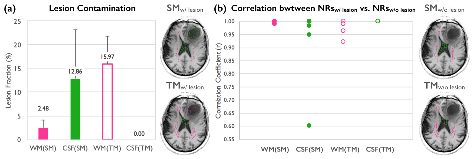
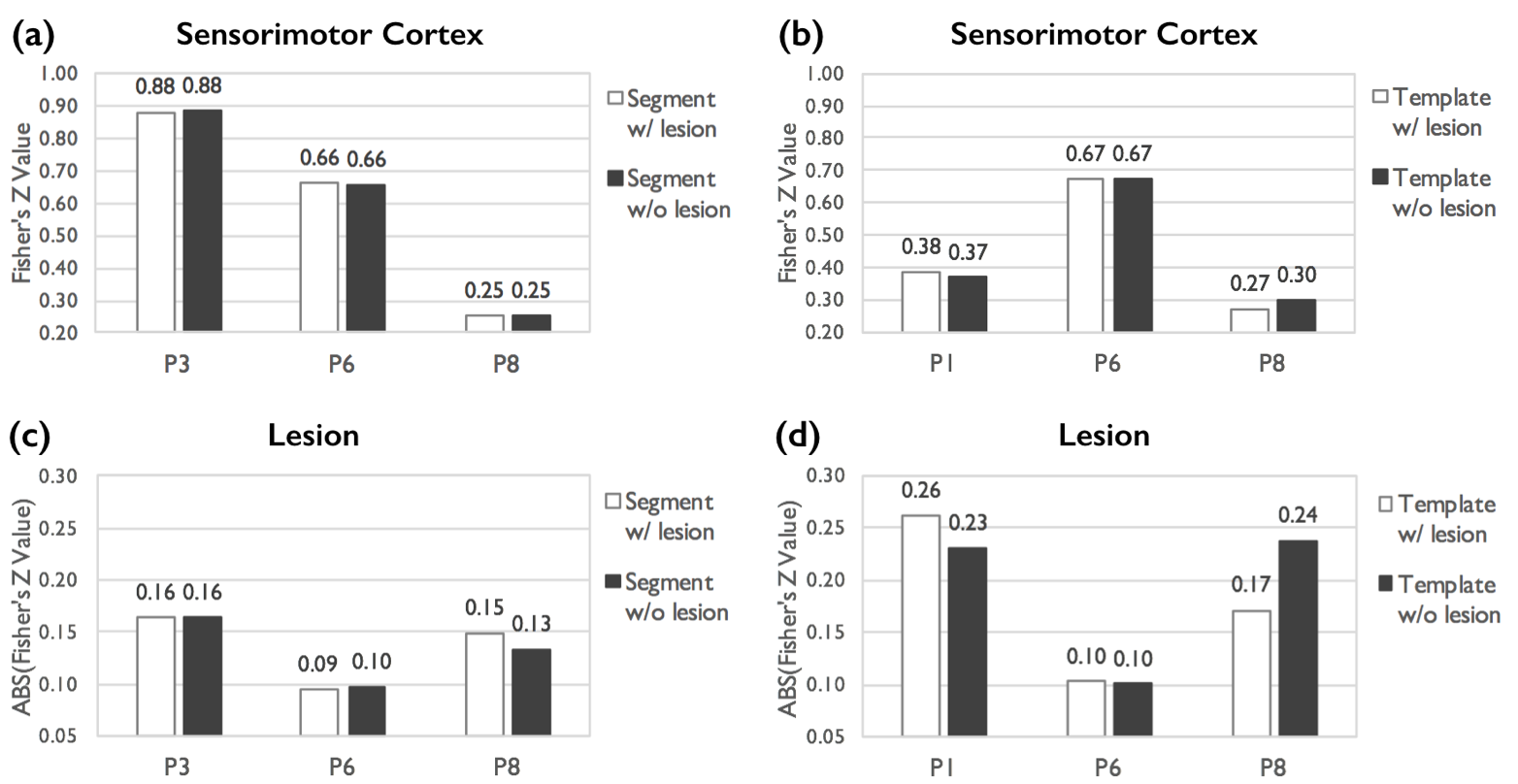
Among the 8 patients, 6 had lesions included in the SMs and 7 in the TMs. Figure 1(a) displays the volume fraction of the lesion in the SM and TM of the WM and CSF. For the SM, lesions had higher chance to be classified as the CSF compared to the WM. In contrast, the lesion had no chance to be classified the CSF in TM. Although the lesion was misclassified to different type of tissues as methods varied, the correlation coefficients between NRs with vs. without the lesion were higher than 0.99 except for three cases each in the segmentation and the template approaches, respectively (Fig. 1b). The connectivity strengths resulted from NR with vs. without lesion contamination were compared in these patients. In the sensorimotor cortex, the connectivity strength (z value) was almost identical among patients in for SMs with and without lesion (Fig. 2a); however, compared to variable results were observed with the TMs (Fig. 2b). In Lesion ROIs, slightly changes in z value were observed in SM with vs. without lesion (Fig. 2c), but larger variations were observed with the TMs. When grouping data from all the patients, no significant difference (p>0.05) were found in connectivity strengths derived with NRs with vs. without lesion contamination for both segmentation and template approaches.CONCLUSION
The WM and CSF masks obtained by both segmentation and template approaches were contaminated by lesion in most of our patients. However, no significant difference in connectivity strength was found due to this effect, which may be related to the small sample size of this study. Comparing to the segmentation approach, more variable results were observed when the template was used for the NR mask determination.Acknowledgements
No acknowledgement found.References
1. Lee MH, Miller-Thomas MM, Benzinger TL, et al. Clinical Resting-state fMRI in the Preoperative Setting: Are We Ready for Prime Time? Top Magn Reson Imaging. 2016(25): 11–18.
2. Jo HJ, Saad ZS, Simmons WK, et al.. Mapping sources of correlation in resting state FMRI, with artifact detection and removal. Neuroimage. 2010(52): 571–582.
3. Song X-W, Dong Z-Y, Long X-Y, et al. REST: a toolkit for resting-state functional magnetic resonance imaging data processing. PLoS ONE. 2011(6): e25031.
Figures