2201
Anticorrelated networks dysfunction in patients with brain tumor in frontal lobeChen Niu1, Xiao Ling1, Pan Lin2, Kun Zhang3, Xin Liu4, Hao Song2, Liping Guo1, Wenfei Li1, Maode Wang5, and Ming Zhang1
1Department of Medical Imaging, The First Affiliated Hospital of Xi'An Jiaotong University, Xi'An, People's Republic of China, 2Key Laboratory of Biomedical Information Engineering of Education Ministry, Institute of Biomedical Engineering, Xi’an Jiaotong University, 3Department of Electronics Engineering, Northwestern Polytechnical University, 4Technical University Munich, 5Department of neurosurgery, The First Affiliated Hospital of Xi'An Jiaotong University, Xi'An, People's Republic of China
Synopsis
The aim of this study is to evaluate whether the lesion in frontal lobe would cause the change of brain positive and negative network interaction, thereby affecting cognitive function. We employed a MSIT to compare the task-positive and DMN changes of brain tumor patients and the healthy controls. Our results indicated the interaction relationship between the task-positive and DMN in patients with brain tumor in the frontal lobe was significantly decreased. We suggest that reduced anti-correlation between DMN and task-positive networks in patients with brain tumor may be caused by dysfunctional DMN, thus affecting the cognitive function of patient.
Introduction:
The anticorrelated network has been suggested as serving to facilitate the coordination and organization of information processing in the brain during the resting and task performance.1 The interactions between task-negative network (DMN) and task-positive network is very important for understanding the intrinsic brain organization.2 Frontal lobe injury often leads to a decline in cognitive function. 3 The aim of this study is to evaluate whether the lesion in frontal lobe would cause the change of brain positive and negative network interaction, thereby affecting cognitive function.Materials and Methods:
Four patients with brain tumors located in frontal cortex and another four age-matched healthy volunteers were enrolled in this study. We used a 3.0T GE scanner equipped with an eight-channel head coil. Both structural images (3D FSPGR 1x1x1 mm3, 140 slices) and BOLD EPI data (TR/TE = 2500/40 ms, flip angle=90°, 3.75x3.75x3mm3) were acquired. All subjects performed a multi-source interference task (MSIT) to activate DMN and the execution/cognitive network (ECN). The detailed information of MSIT is described in reference. 4 The first four volumes were excluded from analysis to allow for initial stabilization of the fMRI signal. The fMRI dataset was analyzed with FSL MELODIC ICA software (www.fmrib.ox.ac.uk/fsl/melodic2/index.html), then motion correction was processed using FLIRT (MCFLIRT). Spatial smoothing was performed at 6-mm FWHM Gaussian kernel to reduce noise. The fMRI images were filtered with high pass filter. The functional images were normalized to the MNI152 standard brain space through their structure images. ICA analysis was carried out using probabilistic independent component analysis (PICA). The number of components was automatically estimated from the fMRI data using a Laplace approximation to the Bayesian evidence of the model order. The resulting maps were thresholded using an alternative hypothesis test based on fitting a Gaussian/Gamma mixture model to the distribution of voxel intensities within spatial maps and a posterior probability threshold of p > 0.5.Results:
The task-positive network and task-negative network (DMN) activation map obtained from healthy controls and patients with brain tumor after MSIT are shown in Figure 1. Compared to healthy controls, we can observe a disturbed relationship between the DMN and task-positive network in patients with brain tumor in frontal lobe. In Figure 2, the results indicate a competitive relationship between brain functional networks of healthy controls, that there’s a negative correlation between spontaneous activity occurring in the default mode network (DMN) and task-positive network. Compared to healthy controls, the negative correlation of tumor patients has reduced, or even showing a positive correlation trend (Patient #4, Fig.2).Discussion:
In this study, we employed a MSIT to compare the task-positive and task-negative network (DMN) changes of brain tumor patients and the healthy controls. Our results indicated that the interaction relationship between the task-positive and task-negative network (DMN) in patients with brain tumor in the frontal lobe was significantly decreased comparing to healthy controls. Some previous studies suggested the DMN was an intrinsic brain system that participates in internal modes of cognition. In addition, the interaction between task-positive network and DMN is closely related with cognitive behavior of the brain. 5 Based on the above results, we suggest that reduced anti-correlation between DMN and task-positive networks in patients with brain tumor in frontal lobe may be caused by dysfunctional DMN, thus affecting the cognitive function of patient. In summary, understanding the interaction between task-positive network and DMN enables us to effectively observe the cognitive changes of patients with brain tumors in frontal lobe, which provides new insights to the preoperative cognitive assessment in clinical practice, and future longitudinal studies of cognitive function abnormality.Acknowledgements
This work was supported by the National Natural Science Foundation of China (61401363, 61262034), by the Fundamental Research Funds for the Central Universities of China (1191320118).References
1. Fox M D, Snyder A Z, Vincent J L, et al. The human brain is intrinsically organized into dynamic, anticorrelated functional networks. Proc Natl Acad Sci U S A. 2005 Jul 5;102 (27):9673-8.
2. Anticevic A, Cole M W, Murray J D, et al. The role of default network deactivation in cognition and disease. Trends Cogn Sci. 2012 Dec;16(12):584-92.
3. Stuss D T. Functions of the frontal lobes: relation to executive functions. J Int Neuropsychol Soc. 2011 Sep;17 (5):759-65.
4. Bush G, Shin L M. The Multi-Source Interference Task: an fMRI task that reliably activates the cingulo-frontal-parietal cognitive/attention network. Nat Protoc. 2006;1(1):308-13.
5. Buckner R L, Andrews-Hanna J R, Schacter D L. The brain's default network. Ann N Y Acad Sci. 2008; 1124(1):1-38.
Figures
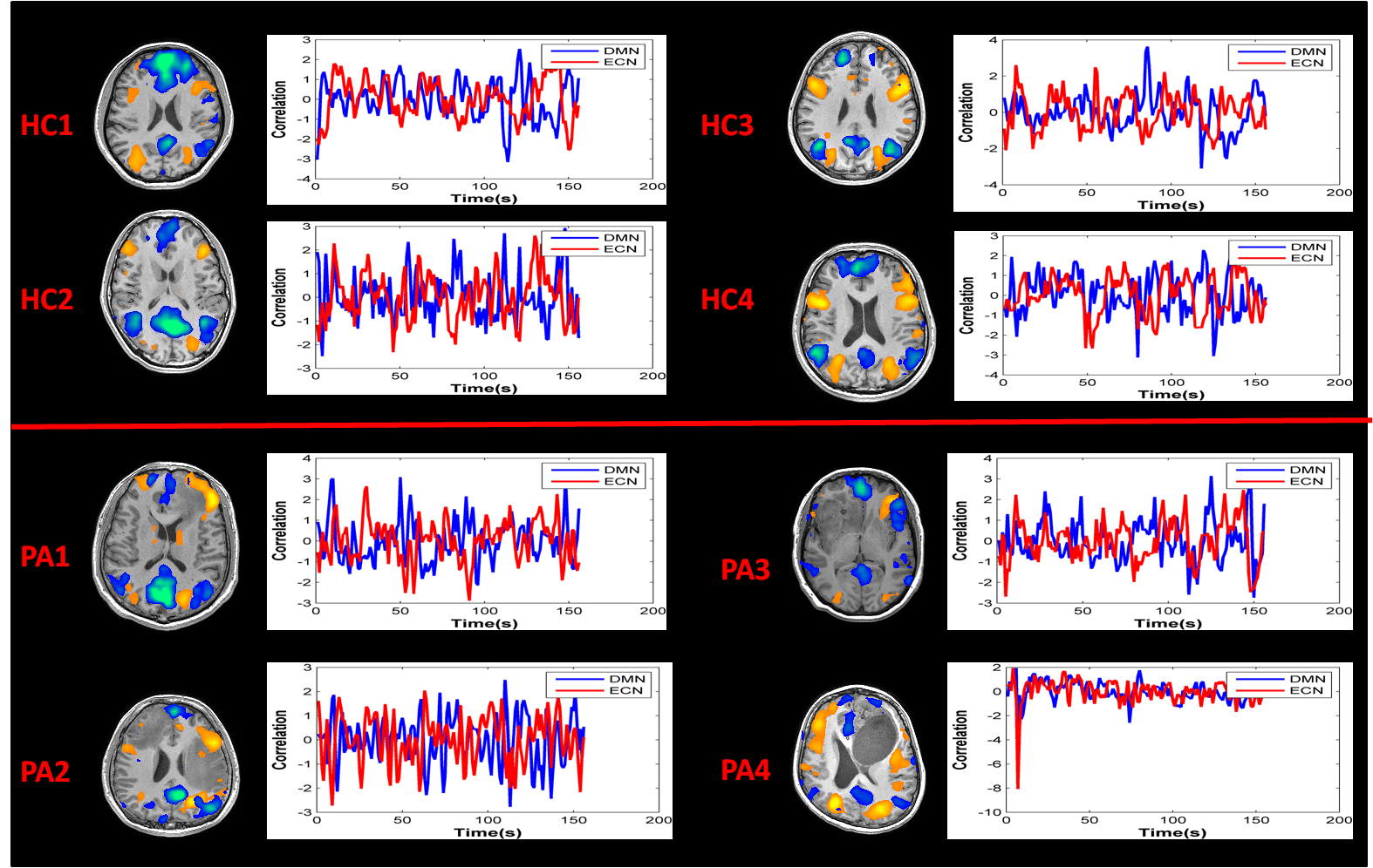
Fig.1 Intrinsic Network and reaction time for each healthy control and patient with brain tumor. The blue curve represents the regions of the task-negative network (DMN), while red curve represents the regions of the task-positive network (ECN).
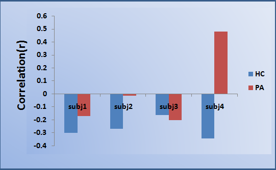
Fig.2. Average correlation values across the task-negative (DMN) and task-positive regions are shown for healthy controls (blue) and the patients with brain tumor (red).