2189
USPIO-enhanced magnetic resonance imaging of N-ethyl-N-nitrosourea- induced endogenous rat gliomas: contribution of USPIO-related iron-loaded microglia to signal changes1Radiology, Shiga University of Medical Science, Otsu, Japan, 2Radiology, Kohka public hospital, Kohka, Japan, 3Shiga University of Medical Science, Otsu, Japan
Synopsis
We assessed whether ultrasmall superparamagnetic iron oxide (USPIO) phagocytosed by microglia contribute to tumor enhancement in MR imaging of N-ethyl-N-nitrosourea (ENU)-induced endogenous rat brain gliomas by comparing MR images with the corresponding histological sections. USPIO-enhanced MR imaging may provide pure vascular bed images in the early phase within 6 hours after intravenous administration of USPIO agent because it was histologically proved that the administrated agent remained within intravascular space. In the late phase at 63-66 hours after the administration, USPIO-enhanced Gradient-Recalled-Echo T2-weighted images may depict the distribution of microglia that were magnetically labeled by USPIO agent.
Purpose
We assessed whether ultrasmall superparamagnetic iron oxide (USPIO) phagocytosed by microglia contribute to tumor enhancement in MR imaging of N-ethyl-N-nitrosourea (ENU)-induced endogenous rat brain glioma model by comparing MR images with the corresponding histological sections.Materials and Methods
Animal model
13 Tumors were induced in 10 Fischer 344 rats by transplacental exposure of litters to ENU. All following experiments were complied with the National Institute of Health’s Guide for the Care and Use of Laboratory Animals, and the Animal Care Guidelines of our institution.
MR imaging
We used a horizontal 7.0-Tesla MR scanner with a superconducting magnet with a 40-cm clear bore and a 12-cm bore actively shielded gradient coil set capable of producing magnetic field gradients up to 400 mT/m. The rats were anesthetized with 4.0 % isoflurane and maintained for 2.0-2.5% isoflurane in 40% oxygen and 60% air through the nose during imaging procedures.
Pre-contrast baseline and gadolinium (Gd)-enhanced MR imaging
Spin-echo (SE) T2-weighted images, SE T1-weighted images, and gradient-recalled-echo (GRE) T2-weighted images were acquired to establish the pre-contrast baseline. Each sequence has 14 images with section thickness: 1.2 mm, intersectional gap: 0.2 mm, field of view: 30 × 30 mm, and matrix size: 256 × 128. Subsequently, rats were intravenously injected with 0.4 mmol/kg body weight of Gd contrast agents and were scanned for contrast-enhanced SE T1-weighted images with the same acquisition parameters as the pre-contrast baseline SE T1-weighted images.
USPIO-enhanced MR imaging
The rats were intravenously injected with 10 mgFe/kg body weight of USPIO agent (Meito Sangyo, Aichi, Japan). The USPIO-enhanced MR images with the same acquisition parameters as the pre-contrast baseline MR imaging set, including SE T2-weighted images and GRE T2-weighted images, were acquired at 5-6 hours after USPIO administration for early phase imaging (6 tumors in 4 rats) and at 63-66 hours for late phase imaging (7 tumors in 6 rats).
Histopathology
Following MR imaging, the rats were sacrificed under deep anesthesia. The brains were removed and sliced for matching the location of histological specimens to those of the MR images. Sections were stained with Prussian blue to localize iron oxide, and cell spots were counterstained with Nuclear Fast Red. In order to detect microglia, specimens were stained with anti-Iba1 antibody.
Analysis
We evaluated whether or not there was regional USPIO enhancement in the tumor area on USPIO-enhanced SE T2-weighted images and GRE T2-weighted images by comparing USPIO-enhanced MR images with the matching pre-contrast MR images. Gd-enhanced MR images were used as a reference (for delineating the tumor area). We determined the number of tumors with USPIO-enhancement, the kind of a signal change (decrease or increase), and the USPIO enhancement area at each phase. We also determined the distribution of iron staining and iron-loaded microglia on histopathological specimens. Next, we evaluated whether or not the location of the iron-loaded microglia on the immunohistochemical sections corresponded to the regional USPIO enhancement in the tumor on the MR images. The signal loss on pre-contrast GRE T2-weighted images were thought to have originated in pre-existent intra-tumor hemorrhage. Two observers reviewed the MR images and the histopathological specimens together.
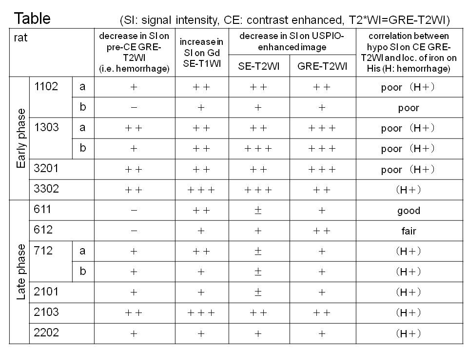
Results
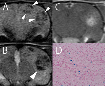
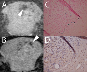
The results are shown in Table. On early phase, the areas with a decrease in signal intensity on SE T2-weighted images are equal to or smaller than the Gd enhancement area in the tumors. GRE T2-weighted images describe the enhancement areas equal to or larger than the Gd enhancement area. GRE T2-weighted images detect a small amount of iron oxide in the late phase. Prussian blue stained iron oxide particles originated from USPIO agent were rarely observed on the histological sections obtained after the early phase imaging (Fig. 1). Histological sections obtained after the late phase imaging revealed iron loaded microglia in the enhancement area on the GRE T2-weighted images (Fig. 2).Discussion
USPIO-enhanced MR images demonstrate strong contrast enhancement in the tumor area in the early phase. Histologically, however, few Prussian blue stained iron oxide particles were observed in the tumor area. It is supposed that most of the administrated agent remained within intravascular space in the early phase and removed by transcardiac perfusion fixation method. Our study also shows histologically that the late phase USPIO-enhanced MR imaging is able to detect intensity changes originated from microglia labeled magnetically by USPIO agent.Conclusion
USPIO-enhanced MR imaging in ENU-induced endogenous rat gliomas is able to provide 1) pure vascular bed images in the early phase within 6 hours after intravenous administration of USPIO agent and 2) distribution of microglia labeled magnetically in the late phase at 63-66 hours after the administration.Acknowledgements
This work was supported by a JSPS KAKENHI Grant Number 21591557.References
No reference found.Figures