2186
Perturbed development of rhesus macaque fetal cerebral cortex and white matter in an IUGR pregnancy, secondary to placental insufficiency, characterized by in utero diffusion MRI1Advanced Imaging Research Center, OHSU, Portland, OR, United States, 2Department of Obstetrics and Gynecology, OHSU, 3Pediatrics, Bioengineering, and Radiology, University of Washington, Seattle, WA
Synopsis
In utero diffusion and anatomical MRI measurements were performed on a naturally occurring intrauterine growth restriction (IUGR) and 3 control pregnant rhesus monkeys. Water diffusion anisotropy (FA) within fetal cerebral cortex as well as white matter was used to characterize abnormal development in the IUGR fetal brain. Markedly higher cortical FA, indicating aberrent morphogenesis of cortical neurons, was observed in the IUGR fetal brain compared to controls. In addition, significantly reduced FA in a number of white matter tracts was also found in the IUGR fetal brain, reflecting perturbed white matter development.
Introduction
Altered brain development is frequently seen in infants born with intrauterine growth restriction (IUGR), the incidence of which is 5% - 7% in all pregnancies1. Recent advances in image reconstruction have made possible high-resolution, in utero measurement of water diffusion anisotropy in the fetal brain2. Thus, in utero diffusion MR has the potential to non-invasively detect abnormal fetal brain development in IUGR pregnancies. In this study, in utero diffusion and anatomical MR measurements were performed on a naturally occurring IUGR and 3 control pregnant rhesus monkeys. Water diffusion anisotropy within fetal cerebral cortex as well as white matter was used to characterize abnormal development in the IUGR fetal brain.Methods
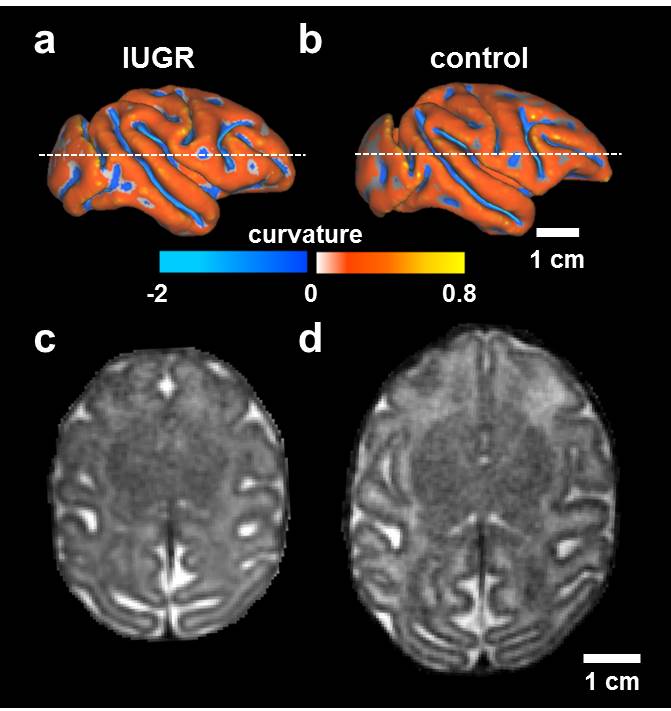
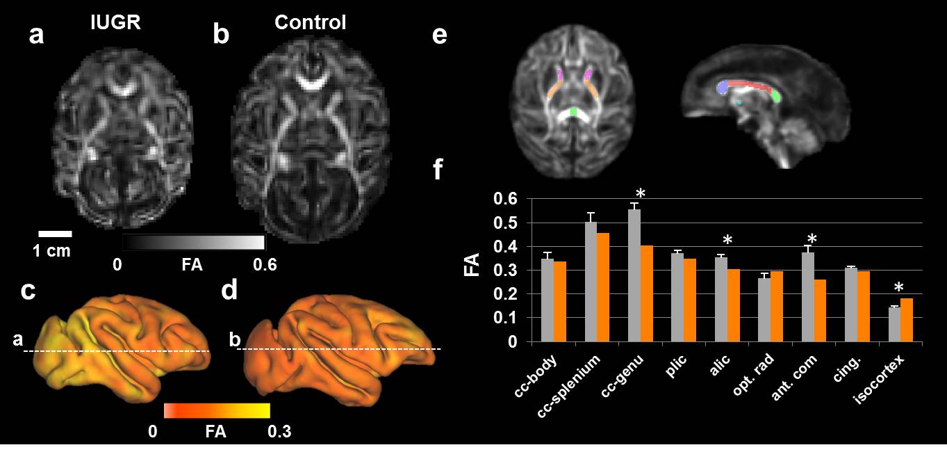
In a cohort of four pregnant rhesus macaques, one was identified as a case of placental insufficiency of unknown cause, as revealed with Doppler ultrasound by reduced uterine artery blood flow. All four pregnant animals underwent in utero MRI examination at gestation day (G)135, of a 168 day term, using a Siemens 3T Tim Trio system. As described previously3, turbo spin-echo (TSE) was used to acquire T2-weighted (T2W) images (TR/TE=5000/97ms) with an in-plane resolution of 0.67mm and thickness of 1mm along the maternal axial, sagittal and coronal axes. An EPI-based diffusion weighted pulse sequence was used to acquire one b0 volume and 20 volumes (b=0.5ms/µm2). Likewise, the diffusion-weighted images were acquired along 3 orthogonal axes at an in-plane resolution of 1 mm and thickness of 3 mm. High-resolution T2W volumes (Figure 1c, d) and FA maps (Figure 2a, b) were reconstructed at isotropic resolutions of 0.5 mm and 0.75 mm, respectively. Fetal brains were manually segmented on the reconstructed T2W volumes using ITK-SNAP. Cerebral cortical surfaces were generated and cortical FA was mapped onto these surfaces using CARET software. Surface curvature, a measure of brain folding, was also computed (Figure 1a, b). An FA template was generated from FA maps of an additional 4 control fetal brains of G135. A label map containing 8 WM regions of interest (ROIs) were manually segmented on this FA template and then transformed to the individual FA map (Figure 2e). Hemisphere and ROI-averaged FA values were reported in this study.Results
The placental insufficiency case was identified as IUGR upon delivery by a markedly smaller fetal weight relative to those of controls (193g vs 333 ± 11g). The fetus of the IUGR case has a smaller brain compared to those of age-matched controls (22 cm3 vs 32 ± 2 cm3) (Figure 1). Despite the similar pattern of cortical folding (Figure 1a, b), the surface area is reduced relative to controls (87 cm2 vs 115 ± 4cm2). Within all characterized WM tracts but the optic radiation, mean FA of the IUGR fetus brain is lower than that of controls (Figure 2e, f), indicating aberrant WM development in the IUGR case4. At the genu of the corpus collosum, anterior commissure, and anterior limb of the internal capsule, FA in the IUGR case was more than three standard deviations (of the control distribution) lower than the control mean (Figure 2f, asterisks). Throughout the cerebral cortical gray matter, FA is higher in the IUGR case compared to controls (Figure 2a-d). This can be appreciated in the lateral views of right hemisphere surfaces of the IUGR brain and a representative control brain (Figure 2c, d), onto which cortical FA is projected. Averaged over entire isocortex, cortical FA in the IUGR brain is 0.182, compared to 0.142 ± 0.006 for the 3 control G135 brains (Figure2c,d,and f). In contrast to WM, higher cerebral cortical FA in the IUGR case signaled less developed neuron dendrites, a perturbed morphogenesis of the cells within developing cerebral cortex relative to controls5.Conclusions
Perturbed development in fetal cerebral cortex and white matter tracts were seen in the IUGR case, indicated by higher cortical FA, and lower white matter FA respectively. In Utero diffusion MRI has the potential to characterize abnormalities in fetal brain development by providing cellular-level information.Acknowledgements
This study was supported by NIH grant R01 AA021981 and grant P51OD011092References
1. Levine et al., Pediatrics, 2015;135(1):126-141
2. Fogtmann et al., IEEE Trans Med Imaging. 2014;33(2):272-289
3. Wang et al., Front Neuroanat. 2015; 9:147
4. Mori et al., Neuron. 2006;51(5):527-539
5. Jespersen et al., IEEE Trans Med Imaging. 2012;31(1):16-32
Figures