2174
The effects of breastfeeding versus formula-feeding on cerebral cortex maturation in infant rhesus macaques1Neuroscience, Oregon National Primate Research Center, Beaverton, OR, United States, 2Advanced Imaging Research Center, Oregon Health and Science University, Portland, OR, United States, 3Casey Eye Institute, Oregon Health and Science University, Portland, OR, United States, 4Food Science and Human Nutrition, University of Illinois at Urbana-Champaign, Urbana, IL, United States, 5Nutritional Sciences, University of Illinois at Urbana-Champaign, Urbana, IL, United States, 6Abbott Nutrition, Columbus, OH, United States
Synopsis
Infant rhesus macaques were studies to test the effects of breastfeeding versus formula-feeding on cerebral cortex maturation. Through analysis of structural and diffusion MR images, brain volume over the first 6 months of life was not significantly altered by formula-feeding versus breastfeeding, or by formula lutein supplementation. However, cellular maturation within cerebral cortical gray matter differed between formula-fed and breastfed animals. Lower gray matter FA of breastfed infants is interpreted to reflect greater neuronal arborization. This difference may be related to the different social experience as well as the nutrient composition of breast milk.
Purpose
Many studies of human infants have shown benefits of breastfeeding over formula feeding for indices of neurodevelopment, including MRI measures of brain maturation1, 2. However, it is not clear whether such findings are due to nutrient content of breastmilk, the maternal-infant interactions of breastfeeding, socioeconomic factors that differ between breastfed and formula-fed infants, or other confounding factors. This study examined MRI measures of brain development in breastfed and formula-fed monkey infants, in which environmental factors can be tightly controlled. In addition it compared formulas providing two different concentrations of lutein and different sources of vitamin E. Lutein is the major carotenoid in human brain and may play an important role in brain development and function3, 4. Among laboratory animal models, only nonhuman primates shares with humans the selective accumulation of lutein in brain4, and therefore provide an appropriate animal model for studies of the role of dietary lutein in brain development. In the developing cerebral cortex, water diffusion anisotropy decreases with maturation as a consequence of increasing complexity in the morphology of neurons5. Here we test the hypothesis that the differing feeding regimens affect maturation of the cerebral cortex, as revealed by cortical diffusion anisotropy.
Methods
In this study, 21 infant macaques were divided into three groups: a breastfed group (2 males; 4 females), a low-lutein formula-fed group (3 males; 4 females), and a high-lutein formula-fed group (4 males; 4 females). In the breastfed group, the mother received a standard laboratory diet (Lab Chow 5047 High Protein Monkey Chow) plus fruits and vegetables. Infants in the low-lutein group were fed a control formula containing 10-20 mcg/L of lutein and synthetic vitamin E (13.3 IU), whereas those in the high-lutein group were fed a formula containing 200 mcg/L of lutein, as commonly found in the breast milk of women consuming a healthy vegetable-rich diet, plus natural α-tocopherol at a concentration matched to the control formula. All infants were scanned in vivo with a Siemens Trio whole body 3T MRI system (Erlangen, Germany) and the MRI data were acquired by a customized primate head 8-channel volumetric array receive coil (Rapid MR International, Columbus, OH, USA) at 4 and 6 months of age. Structural T1-weighted MP-RAGE6 and diffusion tensor imaging (DTI)7 data were acquired. For the latter, 6 images were acquired with b=0, and 25 diffusion sensitization directions were acquired with b=1000 s/mm2. The MP-RAGE images were acquired with a resolution of 0.4 mm isotropic voxels and DTI images with 1 mm isotropic voxels. To provide a common reference space for group-level analysis, the age-specific unbiased T1-weighted brain templates were built from MP-RAGE images of breast-fed subjects and the parcellation of these templates were obtained through registering to inia19 template8. Fractional anisotropy (FA), was calculated for each animal by averaging throughout the set of cortical regions defined in the inia19 template.
Results
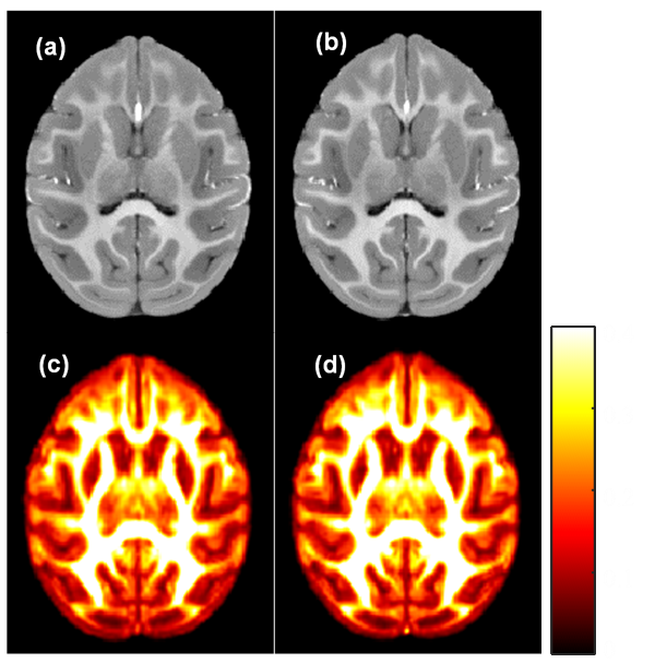
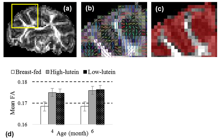
Fig. 1 shows T1-weighted templates and average FA maps derived from breastfed animals at 4 and 6 months. Maturation is reflected in changes in image contrast, particularly within the frontal lobe. A linear model for brain volume and volume/body weight ratio with log transformed age revealed no significant main effect of group nor group by age interaction. As shown in the coronal FA parameter map of a breastfed 6-month-old animal in Fig. 2(a), sizeable diffusion anisotropy is observed within the cerebral cortical gray matter with the data acquisition settings implemented in this study. As is evident in the inset, the primary direction of the diffusion tensor is oriented radially, parallel to the apical dendrites of pyramidal neurons. Repeated measures ANOVA revealed that the mean cortical gray matter FA value in the breastfed group was significantly lower than in both the high- and low-lutein groups (p<=0.05), but no difference was observed between the high- and low-lutein groups. A regional analysis indicates that FA differences are largest in temporal and occipital lobes. These regional effects are currently under more detailed investigation.
Discussion
Brain volume over the first 6 months of life was not significantly altered by formula-feeding versus breastfeeding, or by formula lutein supplementation. Interestingly however, cellular maturation within cerebral cortical gray matter differed between formula-fed and breastfed animals, regardless of formula lutein levels. Lower gray matter FA of breastfed infants is interpreted to reflect greater neuronal arborization. This difference may be related to the different social experience of breastfed versus formula-fed infants as well as the nutrient composition of breast milk. Studies of potential associations between cortical and white matter development are currently underway.
Acknowledgements
This study was supported by Abbott Nutrition through the Center for Nutrition, Learning, and Memory at the University of Illinois, and NIH grant P51OD011092.References
1. Isaacs EB, Fischl BR, Quinn BT, et al. Impact of breast milk on intelligence quotient, brain size, and white matter development. Pediatr Res. 2010;67(4):357-362.
2. Deoni SC, Dean DC, 3rd, Piryatinsky I, et al. Breastfeeding and early white matter development: A cross-sectional study. Neuroimage. 2013;82:77-86.
3. Vishwanathan R, Neuringer M, Snodderly DM, et al. Macular lutein and zeaxanthin are related to brain lutein and zeaxanthin in primates. Nutr Neurosci. 2013;16(1):21-29.
4. McGill TJ, Renner LM, Neuringer M. Elevated Fundus Autofluorescence in Monkeys Deficient in Lutein, Zeaxanthin, and Omega-3 Fatty Acids. Invest Ophthalmol Vis Sci. 2016;57(3):1361-1369.
5. Jespersen SN, Leigland LA, Cornea A, et al. Determination of axonal and dendritic orientation distributions within the developing cerebral cortex by diffusion tensor imaging. IEEE Trans Med Imaging. 2012;31(1):16-32.
6. Mugler JP, 3rd, Brookeman JR. Three-dimensional magnetization-prepared rapid gradient-echo imaging (3D MP RAGE). Magn Reson Med. 1990;15(1):152-157.
7. Basser PJ, Mattiello J, LeBihan D. MR diffusion tensor spectroscopy and imaging. Biophys J. 1994;66(1):259-267.
8. Rohlfing T, Kroenke CD, Sullivan EV, et al. The INIA19 Template and NeuroMaps Atlas for Primate Brain Image Parcellation and Spatial Normalization. Front Neuroinform. 2012;6:27.
Figures