2171
Modulation of structural and functional networks in the ischemic mouse brain by stem cell therapyClaudia Green1, Anuka Minassian1, Andreas Beyrau1, Stefanie Vogel1, Michael Diedenhofen1, Melanie Nelles1, Dirk Wiedermann1, and Mathias Hoehn1,2
1In-vivo NMR, MPI for Metabolism Research, Cologne, Germany, 2Dept. Radiology, Leiden University Medical Center, Leiden, Netherlands
Synopsis
In this study we combined rs-fMRI and diffusion MRI to assess the therapeutic capacity of cortically injected human neural stem cells in the mouse brain after stroke during 3 months. Seed-based analysis of diffusion anisotropy maps and functional connectivity were conducted interrelated with the main focus on the effect of stroke and treatment on the contralesional hemisphere. A delayed breakdown in functional network strength for the therapeutically treated group compared to a sham treated group is observed. However, diffusion anisotropy parameters stay stable with an increasing trend for the thalamus.
Introduction
Combining functional and structural MRI enables a full-fledged view of diverse complex neurological mechanisms and response after therapeutic interventions. In this study we aim to characterize the regenerative capacity of intracerebrally injected human neural stem cells after ischemic stroke in the mouse brain. Repetitive resting-state functional MRI (rs-fMRI) and Diffusion MRI allow longitudinal in vivo characterization of spontaneous or therapeutically induced recovery on a structural and functional level. With our long observation time of 12 weeks after stroke induction we unravel for the first time the long-term effects of cell implantation on structural and functional brain networks.Methods
Rs-fMRI and Q-Ball images (QBI) were acquired before, as well as in week 1, 2, 4, 8, and 12 after ischemic stroke induction. Two days after stroke, stem cells (n=11) / sham (n=6) implantation was carried out in Nu/Nu mice (male, 3-4 months) adjacent to the cortico-striatal lesions. Cell vitality was repetitively measured with Bioluminescence Imaging (BLI). Both MRI scans were always acquired in one session on a preclinical 9.4 T (Bruker, BioSpin) using a cryogenic surface coil with the following parameters and preprocessing steps: rs-fMRI: GRE-EPI sequence; TR/TE=2,840/18ms; 16 slices; slice thickness=0.5mm; matrix=9696; 182x182 µm2 res.; NR = 100. Functional datasets underwent slice-wise motion correction, high-pass filtering (>0.01 Hz), in-plane smoothing (Gaussian kernel=0.3mm) and physiological noise regression of recorded respiratory signals, motion parameters and drifts. QBI: 4-shot SE-EPI; TR/TE = 3,500/20ms; 128x128 matrix; 139x139 µm2 res.; 126 directions + 8 A0 images; Δ = 10ms, δ = 4 ms; b-value= 2,000s/mm2. A0 images were motion corrected and the linear interpolation was applied to the diffusion images. Postprocessing: Seeds of the sensorimotor cortex, as well as the thalamus and caudate putamen (CPu) were extracted after a multi-step process of linear and non-linear co-registrations of the AMBMC mouse brain atlas to the functional and diffusion data. For rs-fMRI, we calculated seed-based full cross-correlation maps with FSLNets (FMRIB Software Library). Diffusion anisotropy maps were generated and fiber tracking was performed with DSI-Studio.Results
Mean z-scored correlation maps of unilateral and bilateral functional connectivity from the stem-cell treated group show a delayed decrease in connectivity only after week 2 of stroke induction (Figure 1). Interestingly, after week 2, the homotopic contralesional side is severely affected up until the end of the experiment, after week 12, for both groups. Maximal loss of inter-seed correlation is observed in week 8 for the cell treated group and in week 4 for the sham group. The time courses of the intra-seed correlations show a gradual decline for all seeds until 8 weeks after stroke for the cell group, whereas in the sham animals the correlation already starts to increase again after 2 weeks (not shown). Diffusion parameters representing the degree of anisotropy in a specified seed stay on a constant level for the caudate putamen and the somatosensory cortex excluding the limb area (Figure 2). However, the anisotropy curve of the thalamus seems to achieve a plateau from week 4 on after stroke for the animals implanted with a sham solution.Discussion
Our results show a delayed decrease of functional connectivity in the sensorimotor network for the stem-cell group, suggesting a stabilizing function of the cells in the first weeks after stroke. This effect did not sustain longer than four weeks and correlates with the loss of cells, monitored with BLI. The persisting, strongly impacted functional network connectivity on the contralesional hemisphere is partially reflected in the similar results of the time courses from the mean intra-seed correlation. This observation indicates a relation between the strength of an intra-seed correlation and the correlation between seeds. The results from the functional datasets illustrate that a homotopic change in functional networks not only affects short-range interactions, but has a large impact on long-range brain correlations as well. The thalamic time courses of the functional results and the anisotropy indices from the diffusion data reveal an inversely proportional behavior, with a functional delay, demonstrating a relation between structural and functional recovery.Conclusion
In this study we present a new mode of action of the therapeutic capacity of stem cell implantation after stroke in vivo on the neuronal circuits, merging the structural and functional point of view. With our results, we show the importance of a global analysis from different angles, and the necessity for long observation times after stroke. Furthermore, we provide new insight into the structure-function relationship in neurodegenerative diseases.Acknowledgements
This work was supported by grants from the EU-FP7 programs TargetBrain (HEALTH-F2-2012-279017) and BrainPath (PIAPP-GA-2013-612360).References
No reference found.Figures
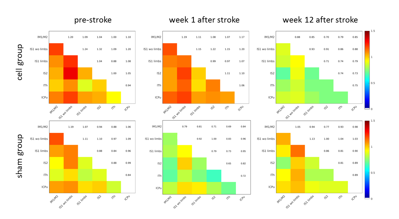
Contralesional
seed-based functional correlation between the cell treated group (upper row)
and the sham animals (bottom row) for the baseline measurement before stroke,
as well as the first and last time point after stroke. The cell treated animals
show a delayed decrease in correlation strength compared to the sham group.
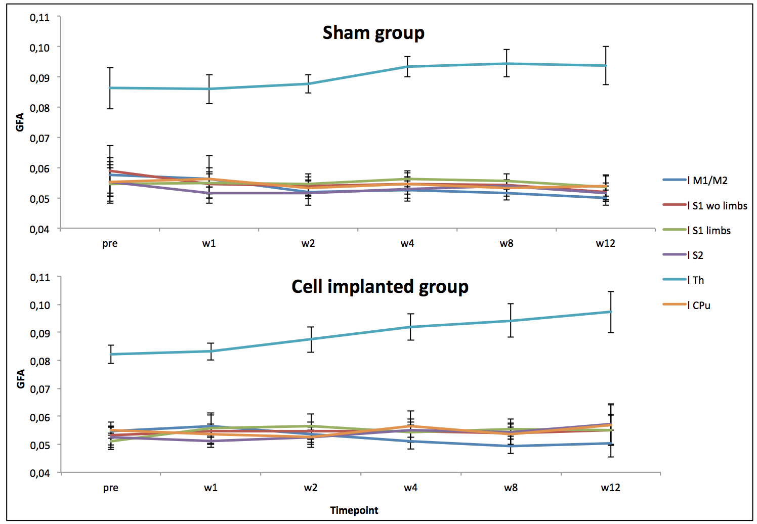
Generalized
fractional anisotropy timecourse for both groups. The
increase in thalamic anisotropy over time is clearly visible. The values for
the sham animals seem to reach a plateau beginning from the fourth week after
stroke, whereas values of the cell implanted group still rise until the end of
the experiment.