2166
The Pain of Pre-Clinical fMRI1Psychology, University of Sheffield, Sheffield, United Kingdom, 2Biological Services, University of Sheffield, Sheffield, United Kingdom, 3School of Clinical Dentistry, University of Sheffield, Sheffield, United Kingdom
Synopsis
This study uses multi-parametric MRI measures to further our understanding on the pathophysiology of migraine in a novel preclinical model of medication overuse headache. Rats undergo sustained triptan exposure inducing cutaneous allodynia. Following drug exposure, we perform ASL, NMR, connectivity and evoked response BOLD fMRI to investigate sustained neuronal adaptations/changes. We investigate how drug overuse leads to latent sensitization to migraine triggers 20 days after exposure. We find significantly altered CBF levels in triptan treated animals. NMR reveals changes in GABA & Tau metabolite levels. fMRI shows changes in functional connectivity and activation of brain regions associated with pain pathways.
Purpose
Migraine is one of the most common debilitating disorders, affecting approximately 16% of the worldwide population. In addition to being a leading cause of pain and suffering, chronic migraine attacks are a source of both high medical costs and loss of productivity. Although evidence indicates that the various phases of migraine involve a number of pathophysiological mechanisms, including inherited alteration of brain excitability; intracranial arterial dilatation; recurrent activation/sensitization of the trigeminovascular pathway; and structural/functional changes in some genetically susceptible individuals1; the neurobiology of migraine is still not completely understood. Hence the current available treatments (be them abortive or prophylactic) are not very effective and often not well tolerated by the patient.
The current study aimed to further our understanding on the pathophysiology of migraine. A clinically relevant model of triptan medication overuse headache in rats2 was used together with multi-parametric pre-clinical functional Magnetic Resonance Imaging (fMRI) to investigate which brain regions were altered under migraine-like conditions. This data alongside behavioural studies allowed investigation of the roles of these changes in the pathophysiology of migraine.
Methods
Naïve rats underwent continuous administration of either Sumatriptan (n=4) or saline (n=4) by subcutaneous osmotic mini-pump (0.6 mg/kg/day). After 7 days the minipump was removed. This pre-clinical rat model mimics the development of migraine symptoms seen in sufferers; i.e. development of cephalic & extra-cephalic cutaneous allodynia following external triggers (sodium nitroprusside - a nitric oxide donor) at day 212,3. Allodynia was assessed using von-Frey apparatus to measure peri-orbital and hindpaw withdrawal thresholds to an applied force.
All fMRI measurements were made at 7 Tesla in a pre-clinical MRI facility (Bruker BioSpec, 310mm bore). RF transmission utilised a 86mm(ID) 1H quadrature volume resonator. Signal reception used a single channel surface coil. Subjects were scanned at the day 0, 7, 14, and 21 time points.
For this in depth longitudinal study rats were anaesthetised with Isoflurane in a 30:70 O2/N2 gas mixture. Temperature and breathing rate were monitored throughout. At each time point we performed i) high resolution (256*256) T2/T2* relaxometry, ii) baseline CBF measurement (single slice 96*96 FAIR-EPI), iii) localised 1H PRESS NMR (TR 2s, TE 24ms, NS=512, 56ul voxel), iv) connectivity fMRI and v) evoked activity Blood Oxygenation Level Dependent (BOLD) fMRI. Evoked activity fMRI utilised standard single shot GE-EPI (64*64, 1s TR, 12ms TE, 90degree flip angle, 9 slices) during either 30s binocular light (5Hz/20Hz white LED illumination) or 16s RHS whisker pad air puff stimulation; with 96s inter stimulus interval to ensure the associated heamodynamics had returned to baseline.
Results
Following removal of the s.c. minipump (day 0), baseline ASL fMRI cerebral blood flow measures in grey matter and mid brain structures were significantly reduced in triptan treated (76.2±3.8 ml/100g/min) versus control animals (97.0±10.6 ml/100g/min) at day 2. This reduction persisted even when behavioural sensory thresholds returned to normal (day 20). During this 'latent sensitization' period and following sodium nitroprusside triggers both groups showed an increase in baseline CBF but the mismatch in level persisted (95.3±7.2 & 131.1±4.2 ml/100g/min in treated and controls respectively).
1H PRESS NMR data was analysed using a Phython based Vespa spectral analysis toolbox using a predefined starting basis set (Gaussian line fitted) for the metabolite of interest. Data from a 56ul voxel covering the thalamus showed a decrease in both GABA and Tau in triptan treated animals.
Connectivity fMRI shows enhanced connections between pain related brain structures in treated vs control groups.
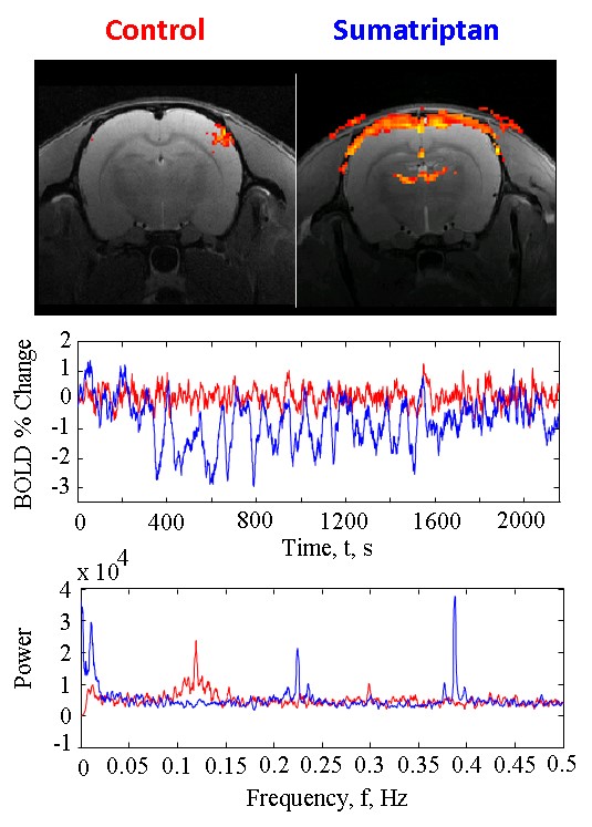
Evoked fMRI data show that triptan-induced latent sensitization is accompanied by enhanced sensitivity to triggers of migraine attack (inc. light/whisker stimulation). Following the stimulation onset (t=60s), BOLD fMRI data show a distinct 0.395Hz oscillation only in areas of superficial cortex and thalamus (Fig 1) (associated pain pathway) in triptan treated rats. The oscillation continues for 1300s and we hypothesise is indicative of systemic pain following the stimulus. It is not observed in control rats. The frequency was not linked to any physiological measure (breathing 0.67Hz/heart rate 0.2Hz). Additionally, control rats showed a distinct V-signal oscillation ~0.1Hz in thalamic areas that is not present in triptan treated rats; counterintuitive given the baseline CBF differences.
Discussion & Conclusion
We carried out a broad range of MR imaging applications to validate a pre-clinical model of migraine. The data from these studies enabled us to correlate MRI changes with the development of increased pain perception and the expression of specific indicators of altered excitability, thus advancing understanding of the mechanisms underlying the development of migraine. The development of this non-invasive preclinical model of migraine will be a useful new tool for the investigation of new anti-migraine treatments.Acknowledgements
MDF was supported by an EU fellowship (PII-GA-2013-626845). All MRI experiments were supported by the faculties of Science and Medicine & Dentistry at the University of Sheffield.References
[1] Noseda R et al. Nat Neurosci.13(2):239-45.
[2] De Felice M. et al. Ann Neurol.67(3):325-37.
[3] De Felice M. et al. Brain.133(Pt 8):2475-88.
Figures