2160
MRI Characterization of Lung Lesions from Pulmonary Tuberculosis1Radiology, Cancer Hospital of Chinese Academy of Medical Sciences at Shenzhen, China, Shenzhen, People's Republic of China, 2Radiology and Imaging Sciences, Emory University School of Medicine, Atlanta, GA, United States, 3MR Technical Services, Siemens Healthcare Ltd., People's Republic of China, 4Radiology, Longhua People’s Hospital of Shenzhen, People's Republic of China
Synopsis
This work demonstrated that lung MRI can be applied to imaging and characterize the abnormalities and lesions in patients with history of pulmonary tuberculosis (TB) with the validation of clinical CT. Results and examples show that MRI not only offers a non-radiation imaging alternative to CT for the lung examination, but also can provide additional information on lung soft tissue properties.
Synopsis
This work demonstrated that lung MRI can be applied to imaging and characterize the abnormalities and lesions in patients with history of pulmonary tuberculosis (TB) with the validation of clinical CT. Results and examples show that MRI not only offers a non-radiation imaging alternative to CT for the lung examination, but also can provide additional information on lung soft tissue propertiesAbstract
Introduction
Given ionizing radiation involved in CT, there is an increased concern on repeated use of CT in patients with tuberculosis (TB), non-cystic fibrosis, pneumonia, and lung cancer, who need follow-up observations. Cautions are also given when using CT for pregnant women, children and adolescents. MRI not only offers a potential non-radiation imaging alternative for radiological examination of lung, but also can provide additional information on the change of soft tissue properties comparing to CT. However, due to the technical challenges in low signal and sensitivity as well as inherited difficulties in motion control, so far, there are very limited reports and clinical experiences in use of MRI for the diagnosis and characterization of abnormalities from pulmonary tuberculosis. Here we report MRI characterizations of lesions from various cases of obsolete pulmonary tuberculosis with a clinically implementable protocol, demonstrating the clinical utility of using MRI to study affected lung of TB patients.
Materials and Methods
Participants: Twenty-five patients with history of pulmonary tuberculosis (16 males, 9 females, mean age of 46 years old) were recruited in this study. All patients were diagnosed TB by the routine clinical examinations for tuberculosis, including chest CT, and had been treated for TB. Written consents were obtained from all participants. Subjects were screened for MRI safety.
MRI scans and Data Analysis: All MRI scans were performed with subject free breathing on a 1.5 T MR scanner (Siemens Avanto) using a body wrap coil with 12 coil elements. Images were recorded using a chest MRI protocol built and optimized for imaging lung, including T1-vibe-dixon, T2-weighted turbo spin echo (TSE) and T2-weighted TSE fat suppressed imaging sequences with imaging parameters of: FOV=360 mm, matrix of 256, TR/TE=3000/60 ms for axial imaging and TR/TE=5000/100 ms for coronal imaging, FA=150 degree. 46 slices with 3 mm slice thickness with no gap was used to cover the lung (resolution: 1.1x1.1x3 mm3). To minimize the respiratory motion artifacts, T2-weighted and T2-weighted fat suppressed coronal images were recorded with parameters of TR/TE=4000/85 ms and 3000/85 ms using a liver dome scout combine with BLADE technique for suppressing motions. The MRI exam was typically 42 minutes. All images were evaluated by two experienced radiologists double-blinded. All lesions found in MRI were confirmed by the clinical standard CT.
Results and Discussions
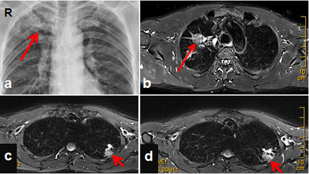
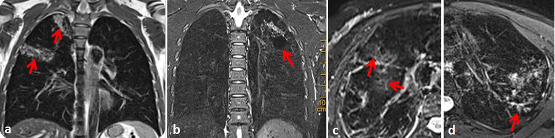
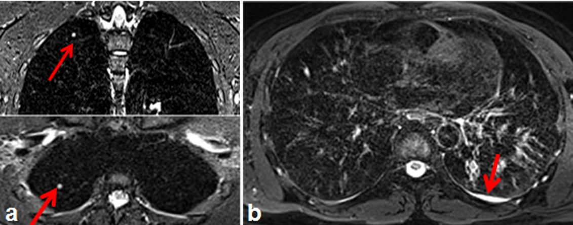
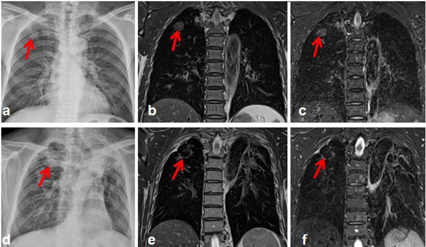
Our results showed that all lesions from TB patients can be visualized by the MRI protocol of this study. Respiratory and other motion artifacts were suppressed by motion control techniques used, resulting high resolution interpretable lung images. The lesions found in MRI can be characterized as: 1) pulmonary nodules (n=12) showing Figure 1a-1d, 2) pulmonary consolidation (n=3) (Figure 2a), 3) pulmonary bulla (n=2) (Figure 2b), 4) ground-glass opacity (GGO) (n=4) (Figure 2c). 5) tree-in-bud sign (n=7) (Figure 2d), and 6) lung interstitial involvement (n=3). A foci with arc and circuitous high signal inside (Figure 2c), which is similar GGO on CT, can be readily identified. With high resolution and soft tissue contrast, MRI was able to detect the small pulmonary nodules (diameter ≤ 5mm) (n=8) in the lung (Figure 3a). In some cases in which the small nodules appear to be heterogeneous, MRI not only delineated the clear boundary of the lesion but also revealed additional features in the nodules, although further pathology examination and correlation of those image features are needed. Using fat-suppressed T2 weighted imaging, lung MRI is sensitive to detect pleural involvement, especially for mild pleural effusion (Figure 3b). Detection of mild pleural effusion (n=7) was 100%. Interestingly, the lung MRI protocol of this study is capable of revealing not only calcified tuberculoma, but also the heterogeneous features of the calcified lesion, while CT may only show homogeneous calcified tuberculoma with high density (n=3) (Figure 4).
Conclusions
MRI was able to provide motion reduced high resolution images of lungs and identify various types of lesions from obsolete pulmonary tuberculosis. The modified T2-weighted fat suppression imaging enables classifying the lesions of small pulmonary nodules, calcified tuberculoma, as well as the heterogeneity of calcification. The current study suggests that clinical implementation of MRI for imaging pulmonary tuberculosis is feasible and potentially offers more diagnostic information than the clinical standard CT.
Acknowledgements
No acknowledgement found.References
1. Montella, S., et al., Magnetic resonance imaging is an accurate and reliable method to evaluate non-cystic fibrosis paediatric lung disease. Respirology, 2012. 17(1): p. 87-91.
2. Montella, S., et al., Assessment of chest high-field magnetic resonance imaging in children and young adults with noncystic fibrosis chronic lung disease: comparison to high-resolution computed tomography and correlation with pulmonary function. Investigative radiology, 2009. 44(9): p. 532-538.
3. Schloß, M., et al., Magnetic Resonance Imaging of the Lung as an Alternative for a Pregnant Woman with Pulmonary Tuberculosis. Journal of radiology case reports, 2015. 9(5): p. 7.
4. Ekinci, A., et al., MRI of pneumonia in immunocompromised patients: comparison with CT. Diagnostic and interventional radiology (Ankara, Turkey), 2016.
5. Ozcan, H.N., et al., Magnetic resonance imaging of pulmonary infection in immunocompromised children: comparison with multidetector computed tomography. Pediatric Radiology, 2016: p. 1-8.
6. Rizzi, E.B., et al., Detection of Pulmonary tuberculosis: comparing MR imaging with HRCT. BMC Infect Dis, 2011. 11: p. 243.
7. Heye, T., et al., Detection and size of pulmonary lesions: how accurate is MRI? A prospective comparison of CT and MRI. Acta Radiologica, 2012. 53(2): p. 153-160.
Figures