2140
Large-scale production of highly-polarized 129Xe1Academic Unit of Radiology, University of Sheffield, Sheffield, United Kingdom
Synopsis
Rapid production of large volumes of highly polarized 129Xe with continuous-flow spin-exchange optical pumping (SEOP) 129Xe polarizers is vital for high-throughput hyperpolarized (HP) 129Xe lung imaging and emergent clinical applications with dissolved 129Xe, e.g. brain perfusion. However, the production rate is limited by cell volume, previously between 300-1500 cm3. Here we present a custom-built 129Xe polarizer designed with a SEOP cell volume of 3500 cm3 which can produce 129Xe polarized to 35% at a Xe production rate of 1200 mL/hour, enabling high-SNR 129Xe lung imaging of naturally abundant Xe and high-SNR 129Xe brain imaging with isotopically-enriched Xe.
Purpose
Rapid production of large volumes of highly polarized 129Xe on continuous-flow spin-exchange optical pumping (SEOP) 129Xe polarizers is challenging due to the flow limitation imposed by the SEOP cell volume as the 129Xe polarization decreases beyond a critical flow rate for which the 129Xe residency time in the SEOP cell is less than the 129Xe-Rb spin-exchange time. Successful employment of a large cell volume (>1500 cm3) requires two main criteria: i) a highly homogeneous static B0 field over the cell volume; and ii) a well-collimated, high-power optical pumping laser beam which is homogenous over the cell length. Presented here is a custom large-scale 129Xe SEOP polarizer featuring a 3500 cm3 SEOP cell which is designed to work in a clinical setting.Materials and Methods
129Xe polarizer main components: Four square coils (100 cm width) with geometry based on the design of Merrit et al.1 for optimized B0 homogeneity at 3 mT; 3500 cm3 (dimensions 7.5 cm diameter, 80 cm length) cylindrical borosilicate SEOP cell filled with < 1 g rubidium located at the SEOP cell gas inlet. 200 W diode in combination with a beam expander to produce a 7.5 cm diameter circular beam; storage field (~0.2 T) and spiral glassware (8 rings, 4.5 cm diameter, 1 cm thickness) for collection of frozen Xe. 129Xe SEOP flow parameters: A gas mixture of 3% Xe, 10% N2, 87% He was flowed through the cell at a volumetric flow rate of 600 sccm (standard cubic centimetres per minute) and collected in its solid state within a cryostat (spiral submerged in liquid nitrogen) for 50 min. The recovered gaseous Xe volume after submerging the spiral in hot water was 1000 mL, where the Xe was collected in pre-evacuated Tedlar bag. The oven housing the SEOP cell was regulated to T = 130 °C and the SEOP cell pressure was ~ 2 atm. These parameters were based on optimized 129Xe polarization from previously reported volume/flow rate production maps.2 The 129Xe polarization was measured after 50 min of cryogenic collection using a polarization method described previously.3 Imaging parameters: Natural abundance Xe (26% 129Xe) lung imaging parameters: 3D SSFP sequence at 3 T, TE/TR = 1.44 ms/4.6 ms; flip angle = 7°; bandwidth = ±8.5 kHz; FOV = 320x320x224mm; voxel size = 4.2x4.2x8 mm. Enriched Xe (86% 129Xe) lung imaging parameters: 3D SSFP sequence at 3 T, TE/TR = 1.38 ms/4.5 ms; flip angle = 10°; bandwidth = ±8.5 kHz; FOV = 320x320x218mm; voxel size = 4.2 mm3. Enriched Xe (86% 129Xe) brain imaging parameters: 2D SPGR at 1.5 T; TR/TE = 34 ms/1. 6 ms; flip angle 12.5; bandwidth = ±2kHz; FOV = 220x220 mm; voxel size = 6.9 mm.Results and Discussion
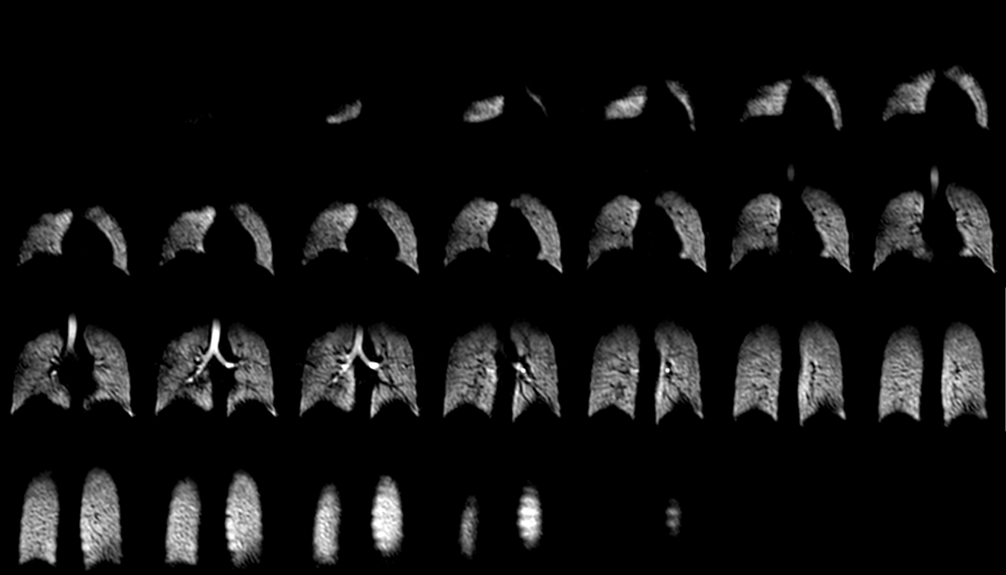
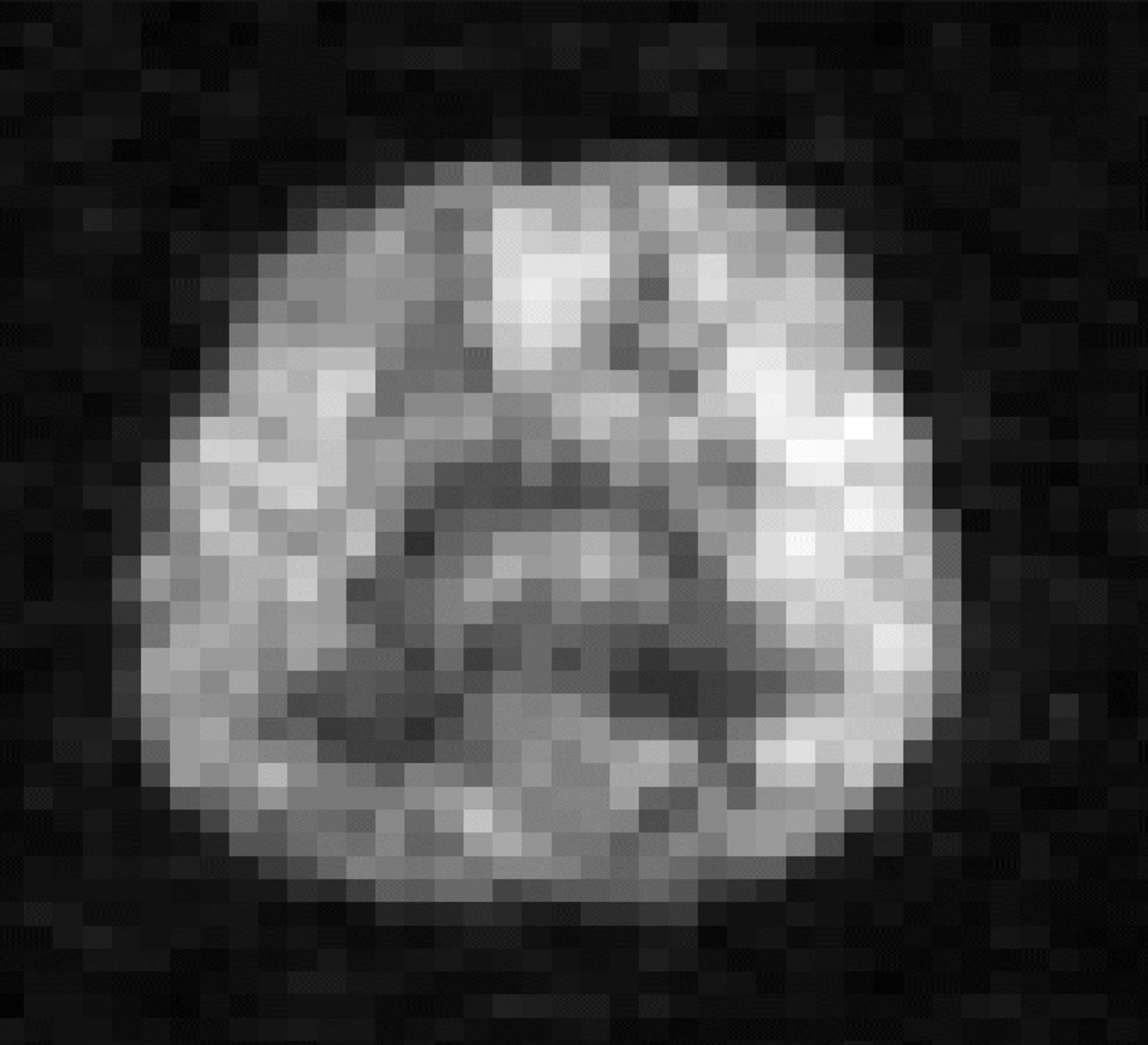
The 129Xe polarization was measured to be 35 % at a Xe production rate of 1200 mL/h. A performance metric called dose equivalence rate has been defined in a previous study as DE rate = f129P129FRXe, where f129 = isotopic fraction of 129Xe (assumed as 1 in the calculation), P129 = 129Xe polarization and FRXe = Xe production rate.3 Using this formula with FRXe = 1200 mL/h and P129 = 35% yields a DE rate = 420 mL/h, considerably higher than those recorded previously in a recent review tabulating 129Xe polarizer performances across the field.3 This high 129Xe polarization value and fast production rate has enabled collection of 1000 mL Xe in 50 min for: i) 129Xe lung imaging with naturally abundant Xe (Fig. 2) with an average image signal-to-noise ratio (SNR) = 80; ii) 129Xe lung imaging with an isotropic voxel resolution of 4.2 mm3 with an average image SNR = 40; and (iii) 129Xe axial brain image with SNR = 30.Conclusion
By optimizing the SEOP cell design and running parameters from previous models, we have demonstrated the generation of highly polarized 129Xe with rapid production rates. With this system, we have demonstrated high-SNR lung imaging with naturally abundant Xe ($25/L) and high-SNR 129Xe brain imaging has been made possible.Acknowledgements
No acknowledgement found.References
1. Merrit, R., et al., Uniform magnetic field produced by three, four, and five square coils. Review of Scientific Instruments, 1983. 54:p.879–882
2. Norquay, G., et al., Approaching the theoretical limit for 129Xe hyperpolarisation with continuous-flow spin-exchange optical pumping. Proceedings of the 23rd Annual Meeting and Exhibition of the International society for magnetic resonance in medicine; 2015, 10-16 May; Toronto, ON, Canada, Program number 1505.
3. He, M., et al., Dose and pulse sequence considerations for hyperpolarized 129Xe ventilation MRI. Magnetic Resonance Imaging, 2015. 33:p. 877-886
Figures