2135
Is ADC heterogeneity helpful in characterizing ductal carcinoma in situ (DCIS) at 3.0T breast MRI1Medical Physics and Research Department, Hong Kong Sanatorium & Hosptial, Hong Kong, Hong Kong, 2Department of Diagnostic & Interventional Radiology, Hong Kong Sanatorium & Hosptial, Hong Kong, Hong Kong, 3Breast Care Center, Hong Kong Sanatorium & Hosptial, Hong Kong, Hong Kong
Synopsis
In this study, we intended to investigate the relationship between ADC heterogeneity of DCIS lesions and DCIS lesioxn morphology, histological grade and BIRADS classification using 3T DW breast MRI. Increasing heterogeneity was observed with increasing DCIS histological grade and increasing BIRADS, but not reaching significance level, was observed based on our results. This study was mainly limited in the small numbers of DCIS lesions, so statistical power has to be further strengthened in future studies with larger sample size.
Purpose
Although ductal carcinoma in situ (DCIS) is a precursor of breast cancer, not all DCIS evolves into invasive breast cancer. Better DCIS categorization is thus important to reduce the possible over-treatment. Diffusion weighted imaging (DWI) is a non-invasive MR technique to assess the tissue structural information without the use of contrast agent. Some studies have reported that decrease in mean apparent diffusion coefficient (ADC) is associated with high grade DCIS [1]. However, mean ADC alone was not able to reflect the tissue structural heterogeneity that might be useful for lesion characterization as reported in literatures for other types of tumors [2]. In this study, we intended to investigate the relationship between ADC heterogeneity of DCIS lesions and DCIS grade and BIRADS classification using 3T DW breast MRI.Methodology
DWI data of histology-confirmed pure DCIS tumors were retrospectively evaluated (29 consecutive female patients with 30 lesions, in which the histological grade of 21 lesions could be retrieved, age: 41±8 years). Prior to DCE-MRI scan, DW images were acquired axially using a fat-suppressed single-shot EPI sequence (TE/TR=102/5800ms, voxel size = 1.8 x 1.8 x 6mm3, b = 0, 1000 s/mm2, averages = 4) at 3.0T MR scanner and a dedicated 4-channel breast coil (Trio, Siemens, Erlangen, Germany). VOIs of DCIS lesions were drawn on the ADC map by referring to DCE-MRI images. The heterogeneity of each tumour VOI was evaluated based on the first degree histogram analysis metrics (skewness and kurtosis) and mean ADC. Wilcoxon rank sum test, with a significance level of 0.05, was performed to assess the association between the (1) mean ADC, (2) skewness and (3) kurtosis and BIRADS classification of DCIS, as well as DCIS histological grade (Van Nuys prognostic index 1: low; 2: intermediate; 3: high). The Wilcoxon rank sum test was also performed to compare the skewness and kurtosis of mass and non-mass DCIS lesions.Results:
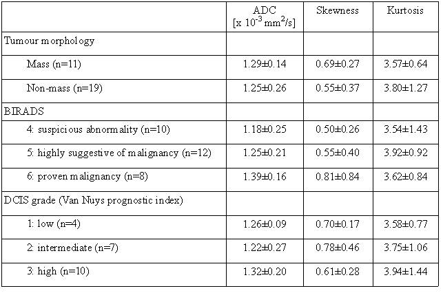
The Patient and lesion characteristics were shown in Table 1. The mean skewness of the DCIS lesion increases with the BIRADS grade (BIRADS 4: 0.50±0.26, BIRADS 5: 0.55±0.40, BIRADS 6: 0.81±0.84), though insignificant (P>0.05). When comparing between different BIRADS grades and the mean kurtosis (BIRADS 4: 3.54±1.43, BIRADS 5: 3.92±0.92, BIRADS 6: 3.62±0.84) and mean ADC (BIRADS 4: 1.18±0.25, BIRADS 5: 1.25±0.21, BIRADS 6: 1.39±0.16) of the DCIS lesion between different BIRADS grades, no significant difference (P>0.05) were observed. When the comparison was performed based on tumor morphology, insignificant difference between mass and non-mass lesions was observed using the mean ADC (mass: 1.29±0.14, non-mass: 1.25±0.26, P>0.05), mean skewness (mass: 0.69±0.27, non-mass: 0.55±0.37, P>0.05) and kurtosis (mass: 3.57±0.64, non-mass: 3.80±1.27, P>0.05). The mean kurtosis value of the DCIS lesion increased with increasing DCIS histological grade (grade 1: 3.58±0.77; grade 2: 3.75±1.06; grade 3: 3.94±1.44), although insignificant difference between DCIS histological grade was found (P > 0.05). However, insignificant difference (P>0.05) was seen when comparing among the mean ADC and mean skewness value of the DCIS lesion of various DCIS histological grades (grade 1: ADC=1.27±0.09, skewness=0.70±0.17; grade 2: ADC 1.22±0.27, skewness=0.78±0.46; grade 3: ADC=1.32±0.20, skewness=0.61±0.28).Discussion
Martincich L et al. concluded that cancer heterogeneity influences the ADC imaging parameters [3]. In this study, increasing heterogeneity was observed with increasing DCIS histological grade and increasing BIRADS, but not reaching significance level. This study was mainly limited in the small numbers of DCIS lesions, so statistical power has to be further strengthened in future studies with larger sample size.Acknowledgements
No acknowledgement found.References
[1] Costantini M, Belliet P, Rinaldi R., et al. Diffusion-weighted imaging in breast cancer: relationship between apparent diffusion coefficient and tumour aggressiveness. Clinical Radiology. 2010; 65(12): 1005-12.
[2] King A, Chow KK, Yu KH, et al. Head and Neck squamous cell carcinoma: diagnostic performance of diffusion-weighted MR imaging for the prediction of treatment response. Radiology. 2013; 266(2): 531-538.
[3] Martincich L, Deantoni V, Bertotto I, et al. Correlations between diffusion-weighted imaging and breast cancer biomarkers. European radiology. 2012; 22(7): 1519-1528.
Figures