2119
Abbreviated Breast Magnetic Resonance Imaging (MRI) for Extent of Disease Evaluation in Newly Diagnosed Breast Cancer1Radiology, UCLA, Los Angeles, CA, United States, 2UCLA
Synopsis
In this retrospective study, an abbreviated protocol consisting of pre-contrast T1 and first post-contrast T1 sequences with fat saturation had near perfect detection of index and secondary cancers, as well as suspicious axillary and internal mammary lymph nodes, in women with newly diagnosed breast cancer. In conjunction with clinical histories and prior imaging examinations, an abbreviated breast MRI protocol is adequate for ipsilateral extent of disease and contralateral breast screening in newly diagnosed breast cancer.
Introduction
Magnetic resonance imaging (MRI) is the most sensitive imaging modality for detecting breast cancer (1). However, access to this powerful imaging modality is limited due to its high cost and relatively long examination time. A recent prospective study proposed an abbreviated scanning protocol in the screening population in order to reduce scan time and interpretation time, thereby potentially reducing cost and increasing access (2). Several recent retrospective studies investigated the diagnostic performances of various abbreviated protocols in newly diagnosed unicentric breast cancer (3-6). All of these studies have shown similar sensitivity, specificity, positive predictive value, and negative predictive value for cancer detection compared to full protocols, yet the authors have only advocated for its use for screening. The purpose of this retrospective study was to compare the diagnostic performance of an abbreviated protocol for extent of disease evaluation and contralateral breast screening in newly diagnosed breast cancer, with the hypothesis that the abbreviated protocol would have similar sensitivity as the full protocol.Methods
In this IRB-approved, HIPAA-compliant, retrospective study, institutional databases were searched for consecutive women with newly diagnosed breast cancer on core needle biopsy who underwent pre-treatment MRI on a 3.0 Tesla scanner at our institution between January 2013 and December 2013. The following selected sequences were interpreted by two fellowship-trained breast radiologists with an average of four years of experience in breast MRI: pre-contrast axial T1 with fat saturation, first post-contrast axial T1 with fat saturation obtained 90 seconds after intravenous contrast injection, and post-processed maximum intensity projection and subtraction images. The radiologists were aware that studies were performed for extent of disease evaluation but were blinded to patient identifying information, demographic information, clinical history, and prior imaging examinations. For each MRI, the presumed index cancer was identified, as well as any additional suspected ipsilateral and/or contralateral secondary cancers. For suspected secondary cancers, a breast imaging reporting and data system (BIRADS) score was assigned. BIRADS 3 was considered negative, and BIRADS 4 and 5 were considered positive. Although not available at the time of image interpretation, whether the radiologists thought a T2 weighted sequence and kinetic information based on additional post-contrast sequences would be helpful was documented. The presence of suspicious axillary and internal mammary lymph nodes was recorded. These interpretations were compared to clinical reports as the reference standard, with histologic confirmation or at least two years of clinical or imaging follow-up in all cases. Sensitivity, specificity, positive predictive value (PPV), and negative predictive value (NPV) were calculated for overall cancer detection of the abbreviated protocol.Results
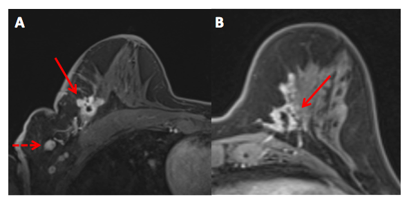
81 women with 92 biopsy-proven breast cancers were included for analysis (mean age 56 years, range 32-92 years). The average cancer size was 26 mm (range, 4 mm-110 mm). For overall cancer detection, sensitivity was 96.7% (89/92), specificity was 25.0% (1/4), PPV was 96.7% (88/92), and NPV was 25.0% (1/4). 100% (19/19) of suspicious axillary lymph nodes and 100% (4/4) of suspicious internal mammary lymph nodes were correctly identified. In 9 women with more than one site of cancer, 95.5% (21/22) of additional sites of disease were detected, seven of which were undetected on prior imaging. Of these seven secondary cancers, six were ipsilateral, and one was contralateral. The one undetected secondary cancer was non-mass enhancement in the contralateral breast. The three undetected index cancers had an average lesion size of 24 mm and consisted of two cases of intermediate grade DCIS and one case of grade 1 invasive ductal carcinoma (IDC) with tubular features, all of which showed progressive enhancement that was subtle on the first post-contrast sequence. One case of DCIS was contralateral to a known index cancer, and the case of IDC was contralateral to a false positive mass containing a microclip, which was incorrectly identified as the index cancer. The three false positive lesions assigned as BIRADS 4 or 5 were three masses. Two of these were in the contralateral breast, and one was in the ipsilateral breast. A T2 weighted sequence and kinetic information based on additional post-contrast sequences were thought to have been helpful in all but one these cases, in which a biopsy-proven fibroadenoma containing a microclip was incorrectly identified as the index cancer.Conclusion
In conjunction with clinical history and prior imaging examinations, an abbreviated breast MRI protocol is potentially adequate for ipsilateral extent of disease evaluation and contralateral breast screening for newly diagnosed breast cancer patients.Acknowledgements
The authors thank Shota Yamamoto, M.D. for his help in study design.References
1. Kuhl CK. MR imaging for surveillance of women at high familial risk for breast cancer. Magn Reson Imaging Clin N Am. 2006;14(3):391-402, vii.
2. Kuhl CK, Schrading S, Strobel K, Schild HH, Hilgers RD, Bieling HB. Abbreviated breast magnetic resonance imaging (MRI): first postcontrast subtracted images and maximum-intensity projection-a novel approach to breast cancer screening with MRI. J Clin Oncol. 2014;32(22):2304-10.
3. Heacock L, Melsaether AN, Heller SL, et al. Evaluation of a known breast cancer using an abbreviated breast MRI protocol: Correlation of imaging characteristics and pathology with lesion detection and conspicuity. Eur J Radiol. 2016;85(4):815-23.
4. Mango VL, Morris EA, David Dershaw D, et al. Abbreviated protocol for breast MRI: are multiple sequences needed for cancer detection? Eur J Radiol. 2015;84(1):65-70.
5. Grimm LJ, Soo MS, Yoon S, Kim C, Ghate SV, Johnson KS. Abbreviated screening protocol for breast MRI: a feasibility study. Acad Radiol. 2015;22(9):1157-62.
6. Jain M, Jain A, Hyzy MD, Werth G. FAST MRI breast screening revisited. J Med Imaging Radiat Oncol. 2016.
Figures