2101
Value of DWI and dynamic contrast-enhanced MRI in differentially diagnosing stage-Ⅰa endometrial carcinomas and endometrial polyps1MRI Division, the First Affiliated Hospital of Zhengzhou University, Zhengzhou, People's Republic of China
Synopsis
Yuan Chen,female,graduated from Zhengzhou University,Master's degree in reading at the First Affiliated Hospital of Zhengzhou University.
Objective
To assess the value of DWI and dynamic contrast-enhanced (DCE) MRI in differential of stage-Ⅰa endometrial carcinomas and endometrial polypsMethods
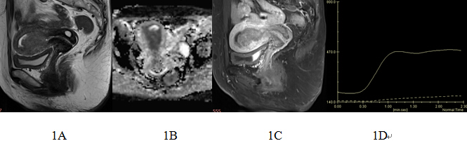
A total of 60 stage-Ⅰa endometrial carcinomas and 38 endometrial polyps confirmed by pathology were analyzed retrospectively. All of patients underwent DWI and DCE-MRI (Siemens Skyra 3.0T MR). The apparent diffusion coefficient (ADC) value, relative apparent diffusion coefficient (rADC) value, time to peak (TTP), maximum contrast enhancement ratio (MCER), the difference value of contrast enhancement ratio between 90s and 60s (ER90s-60s) and time–intensity curves (TIC) type were measured and compared between stage-Ⅰa endometrial carcinomas and endometrial polyps. The diagnostic efficacy of DWI,DCE-MRI in distinguishing stage-Ⅰa endometrial carcinomas and endometrial polyps were calculated.Results
The ADC value ([0.76±0.17]×10-3mm2/s),rADC value(0.58±0.07), TTP([76.47±13.37]s),MCER([119.48±42.51]%)and ER90s-60s([7.12±14.15]%) of 60 stage-Ⅰa endometrial carcinomas were statistically different from 38 endometrial polyps([1.33±0.20] ×10-3mm2/s, [1.02±0.13], [101.86±14.62]s,[ 178.32±88.24]%, [44.67±27.99]%). The cut-off value of ADC, rADC, TTP, MCER and ER90s-60s were respectively 0.904×10⁻³mm2/s, 0.74, 81.5s, 159.01% and 19.25% in differential diagnosis of stage-Ⅰa endometrial carcinomas and endometrial polyps. The sensitivity were 100%,100%,96.43%,53.57%,92.86% and the specificity were 98.31%,100%,93.33%,84.44% and 82.22% for respectively. There were 26 Ⅰ, 31 Ⅱtype TICs and 3 Ⅲtype TICs in 60 stage-Ⅰa endometrial carcinomas. There were 9Ⅱtype TICs and 29 Ⅲtype TICs in endometrial polyps. There was statistically significant difference between the 2 groups.Conclusion
DWI and DCE-MRI are of high diagnostic value in differential of stage-Ⅰa endometrial carcinomas and endometrial polyps. The ADC value, rADC value, TTP, MCER, and ER90s-60s are effective quantitative parameters.Acknowledgements
This research was supported by grants from Key scientific and technological project of Henan province (112102310703).We sincerely thank the patients for their participation, and all the medical staff involved in specimen collecting.
References
[1] Creasman W. Revised FIGO staging for carcinoma of the endometrium. Int J Gynaecol Obstet, 2009,105(2):109.
[2] Tamai K, Koyama T, Saga T, et al. Diffusion-weighted MR imaging of uterine endometrial cancer. J Magn Reson Imaging, 2007,26(3):682-687.
[3] Namimoto T, Awai K, Nakaura T, et al. Role of diffusion-weighted imaging in the diagnosis of gynecological diseases. Eur Radiol, 2009,19(3):745-760.
[4] Wang J, Yu T, Bai R, et al. The value of the apparent diffusion coefficient in differentiating stage IA endometrial carcinoma from normal endometrium and benign diseases of the endometrium: initial study at 3-T magnetic resonance scanner. J Comput Assist Tomogr, 2010,34(3):332-337.
Figures