2095
Can MR quantitative fat fraction technique evaluates bone marrow toxicity during radiotherapy and chemotherapy?1Xijing Hospital, Xi 'an, People's Republic of China, 2GE Research China, GE Healthcare, People's Republic of China
Synopsis
Bone marrow toxicity is very common side effect during radio-chemotherapy treatment of pelvic tumors. In this study, six patients with cervical cancer were included. The Bone marrow fat faction of the subjects were evaluated using quantitative fat fraction MR technique before each week’s treatment and at the end of whole five weeks’ therapy. The results indicated that MRI was sensitive to marrow composition changes and can evaluate the real time bone marrow toxicity during radio-chemotherapy. This could potentially benefit patient with a more optimized treatment plan.
Introduction
Pelvic malignancy is a common malignant tumor, and has a tendency to young people. Concurrent chemo-radiation therapy is often the standard treatment for pelvic malignancies(1). But the side effect which decrease red marrow while increase fatty yellow marrow, results a higher proton density fat fraction(2) (PDFF). This change can reduce hematopoietic capacity and may contribute to bone mineral loss and increased fracture risk in cancer patients(3,4). The purpose of this study is to assess the possibility of real time bone marrow toxicity evaluation during radiotherapy and chemotherapy using MRI based spatial resolved fat fraction technique.Method
This on-going study was approved by the Ethics Committee, and informed consents were obtained from all participants. 6 female subjects (mean age 49 years; age ranging 39 – 60 years) were enrolled in this study. All of the subjects were diagnosed to have cervical squamous cell carcinoma (FIGO IIB-IIIB). Pelvic irradiation dose of subjects was 50 Gy / 25 times, five times a week, Chemotherapy regimen was cisplatin single-agent 40 mg/m²/ week.
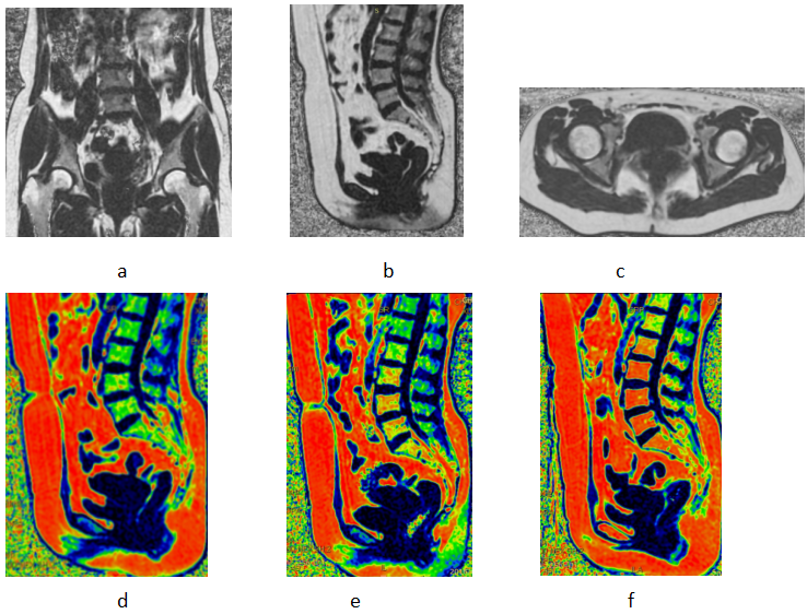
MR scans were performed on a 3.0T MR system (MR750, GE Healthcare, USA) before each week’s treatment and at the end of whole five weeks’ therapy. Commercially available scanning sequence IDEAL-IQ (iterative decomposition of water and fat with echo asymmetry and least-squares estimation quantitation sequence) was used to generate quantitative fat fraction. Detailed scan parameters are as follow: 38cm×38cm field of view, 5mm slice thickness, flip angle 3, matrix 160×160, TR 6.7ms, number of shot 2, 3 TEs per shot, receiving bandwidth 83.3KHz, number of averaging 2. Sequence was scanned in axial sagittal and coronal plane consecutively. ROIs were manually placed on L4 and S1 vertebral body and subcutaneous adipose on the fat fraction images by an experienced radiologist. Final fat fraction was calculated by averaging of the value from 3 planes (Figure 1 a~c).
Results
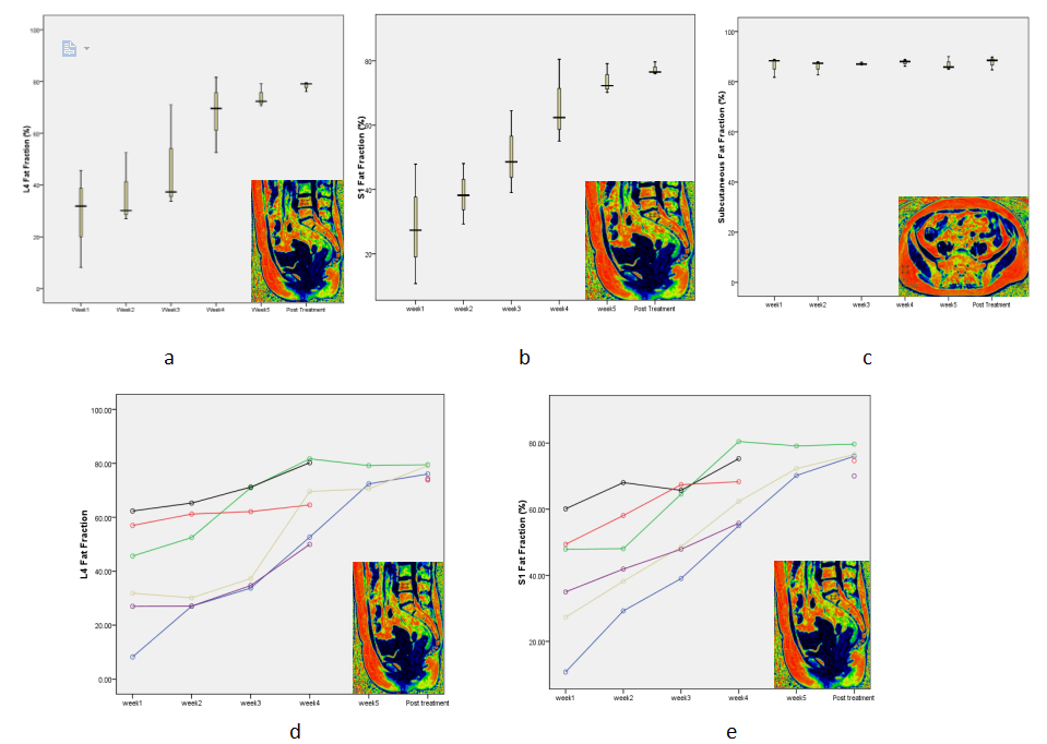
Though out the whole treatment, each subjects demonstrated significant PDFF change. Figure 1 (d~f) shows a subject’s reformatted sagittal PDFF color images at the timing of pre-treatment, three weeks during the therapy and the post-treatment respectively. One can tell the PDFF increases progressively in PDFF throughout all marrow regions, suggesting a systemic conversion of hematopoietic red marrow to more fatty yellow marrow. The time course of treatment changes during therapy can be seen in Figure 2. During the therapy, the PDFF values are respectively 38.66±20.27, 43.89±17.83, 51.66±18.32, 66.46±13.41, 74.04±4.54, 76.58±2.63 in L4 vertebrae, 38.40±17.79, 47,42±13.92, 55.53±11.89, 66.21±10.39, 73.88±4.68, 75.40±3.51 in S1 vertebrae, 85.56±3.29,85.80±3,21,85.44±2.51,85.77±3.49,86.92±2.67,86.78±3.66 in subcutaneous adipose. While the PDFF values in subcutaneous adipose appear stable over time, the PDFF in the L4 and S1 vertebral indicates progressively increasing fat content during the therapy.Conclusion
MR quantitative fat fraction imaging was found to be sensitive to marrow composition changes due to chemoradiotherapy and can evaluate the real time bone marrow toxicity during treatment. Spatially evaluation of toxicity could potentially benefit patient with a more optimized treatment plan(1).Discussion
The results of this study proved that MR quantitative fat fraction technique can sensitively demonstrate bone marrow composition change due to toxicity induced by radiotherapy and chemotherapy. Based on the fat fraction MR images, radiation doctors can adjust radiotherapy plan during treatment accordingly to alleviate toxic marrow from deeper side effect. Both of the 2 subjects whose initial fat faction >50% before treatment experienced stage 3 bone marrow suppression, suggesting a higher possibility of suppression for patients with high initial PDFF. More data will be needed to verify this assumption. Our study also had several limitations. Firstly, the sample size was relatively small, and follow-up time is too short. Secondly, this study did not control for age and menopausal status, which are known to have impact on marrow composition and may confound these results. However, the magnitude of menopausal status and age-related increase in marrow fat is substantially smaller than the treatment effect size seen here (5).Acknowledgements
No acknowledgement found.References
1.Yun L, Bydder M, Yashar C M, et al. Prospective Study of Functional Bone Marrow-Sparing Intensity Modulated Radiation Therapy With Concurrent Chemotherapy for Pelvic Malignancies[J]. International Journal of Radiation Oncology Biology Physics, 2013, 85(2):406–414.
2.Patrick J. Bolan PhD, Luke Arentsen BS, Thanasak Sueblinvong MD, et al. Water–fat MRI for assessing changes in bone marrow composition due to radiation and chemotherapy in gynecologic cancer patients[J]. Journal of Magnetic Resonance Imaging, 2013, 38(6):1578–1584.
3.Rosen CJ, Ackert-Bicknell C, Rodriguez JP, Pino AM. Marrow fat and the bone microenvironment:developmental, functional, and pathological implications. Crit Rev Eukaryot Gene Expr. 2009,19:109–124.
4.Carmona R, Pritz J, Bydder M, et al. FAT COMPOSITION CHANGES IN BONE MARROW DURING CHEMOTHERAPY ANDRADIOTHERAPY[J]. International Journal of Radiation Oncology Biology Physics, 2014, 90(1):155-163.
5.Kugel H, Jung C, Schulte O, Heindel W. Age- and sex-specific differences in the 1H-spectrum of vertebral bone marrow. J Magn Reson Imaging. 2001; 13:263–268.
Figures