2080
Differential Diagnosis of Ovarian Tumor and Degenerated Subserous Leiomyoma Using Diffusion-tensor Imaging1Department of Radiology, the First Affiliated Hospital of Dalian Medical University, Dalian, People's Republic of China, 2GE healthcare China, Beijing, People's Republic of China
Synopsis
Ovarian cancer is one of the most common malignant tumor of female reproductive organs, which is the first cause of death in gynecological malignancies. A mixed cystic and solid appearance of an ovarian mass is usually difficult to differentiate from degenerated subserous leiomyoma. In this study, DTI MR measurements were performed to investigate the difference of the ADC and FA values in ROIs of the soild component region between ovarian tumors and uterine fibroids.
Introduction
Ovarian cancer is one of the most common malignant tumor of female reproductive organs, which is the first cause of death in gynecological malignancies. A mixed cystic and solid appearance of an ovarian mass is usually difficult to differentiate from degenerated subserous leiomyoma. There are many different kinds of ovarian tumors, which show complex histological and morphological types. These may lead to difficulties for the localization and qualitative diagnosis of ovarian tumors. Also due to its asymptomatic nature and deep location in the pelvis, ovarian cancer is often diagnosed at late stages . Therefore, timely and accurate diagnosis is important for treatment and prognosis. Diffusion tensor imaging (DTI) technique has been reported in uterine abnormalities in pelvic MRI, 1 but its use in Ovarian cancer has not been explored. In this study, The apparent diffusion coefficient (ADC) and fractional anisotropy (FA) values of ovarian tumors and degenerated subserous leiomyoma are comapred, to explore the value of DTI in the differential diagnosis of the origin of adnexal masses.Methods
15 patients (53±19.9 years, age range: 8 to 84 years) with mixed cystic-solid ovarian tumors in which solid component accounted for more than 50% were enrolled in this retrospective study. Ethical approval was obtained. The tumor types contain 4 cases of ovarian clear cell carcinoma, 6 cases of serous adenocarcinoma, 1 case of mucinous adenocarcinoma, 1 case of dysgerminoma, 1 case of granular cell tumor, 1 case of malignant germ cell tumor and 1 case of carcinosarcoma. Another 15 patients (45±7.4 years, age range: 36 to 61 years) with degenerated subserous leiomyoma that are difficult to diagnose with conventional MRI were also enroled. All cases were surgically and histologically proved. All the patients underwent MRI exams on a 1.5T scanner (GE Signa HDxt, USA) in a protocol containing the routine T1WI, T2WI, contrast-enhanced MRI, as well as DTI (b =0, 600 s/mm2, in 6 directions). The MR images were blindly reviewed and analyzed by two experienced radiologists, and the values of ADC and FA of the focus were measured using the Functool post-processing software on GE ADW 4.4 workstation(GE, USA). Three regions of interest (ROIs) at the solid sections of the lesions were averaged for final measurements (figure 1) . Each ROI ranged from 50 to 300 mm2. The size of the lesions were also measured. The ADC and FA values of DTI were statistically analyzed using software SPSS17.0 with P<0.05 considered statistical significant. The receiver operating characteristics (ROC) curve was used to evaluate the diagnostic performance.Results
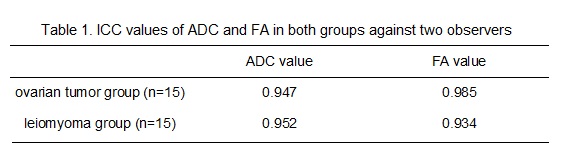
The diffusion tensor images, ADC and FA maps of typical ovarian cancer and subserous leiomyoma patients are shown in Figure 1. The inter-observer agreement was very good. The Intraclass correlation coefficient (ICC) values of ADC and FA in both ovarian tumor group and leiomyoma group are shown in table 1. There was no statistical significance difference of the mean diameter of lesions between two groups. The mean ADC value of the solid sections in the ovarian tumor group (1.188±0.271×10⁻⁹ m2/s) was lower than leiomyoma group (1.497±0.233×10⁻⁹ m2/s) (P<0.05). The mean FA of the solid sections in the ovarian tumor group (0.171±0.055) were significantly lower (p<0.05) than leiomyoma group (0.264±0.046). The cut-off value for FA value was 0.210. The sensitivity and specificity were 93.3% and 73.3%, respectively. The area under the ROC curve was 0.740.Discussion and conclusion
Previous study on ovarian epithelial tumors has reported that there was a statistically significant difference in ADC values between benign and malignant ovarian tumors.2 Mixed cystic-solid ovarian tumors are usually malignant, however uterine leiomyoma are the most common benign tumors in female pelvic tumors. Hence the lower ADC values in ovarian tumors observed in our study is consistent. FA values show the fraction of anisotropic diffusion to total diffusion. The higher the FA, the greater the anisotropy of organization or better directionality. The mean FA value of leiomyoma was seen to be higher in our study. The reason may be that solid portion of degenerated subserous leiomyoma is similar to uterine smooth muscle cells, with closely arranged cell structure. The movement of water molecules is in the same direction, which lead to the higher FA value.
Hence DTI has differential diagnosis value of ovarian tumors and subserous leiomyoma which are difficult to differentiate using conventional MRI sequences.
Acknowledgements
No acknowledgement found.References
1. Kara Bozkurt D, Bozkurt M, Nazli MA, et al. Diffusion-weighted and diffusion-tensor imaging of normal and diseased uterus. World J Radiol, 2015, 7(7): 149-156.
2. Wang YX, Yuan MZ, Wen ZX. Application of apparent diffusion coefficient and exponent apparent diffusion coefficient values in magnetic resonance imaging diffusion-weighted imaging to differentiate benign and malignant ovarian epithelial tumors. J Cancer Res Ther, 2016, 12(1): 401-405.
Figures