2073
Assessment of intravoxel incoherent motion diffusion-weighted MR imaging in solitary pulmonary nodules : comparison and correlation with dynamic contrast-enhanced MR imaging.1Radiology, Tongji Hospital, Huazhong University of Science and Technology, Wuhan, People's Republic of China, 2Radiology, Tongji Hospital of Huazhong University of Science and Technology, Wuhan, People's Republic of China
Synopsis
The study aim to compare the intravoxel incoherent motion (IVIM) and DCE-MRI for distinguishing benign pulmonary nodules and lung cancer and evaluate the diagnostic performance of two methods. We found ADCtotal,D,D*from IVIM and Tmax,SLE from DCE-MRI valuable for differential diagnosis ,with D and Tmax have better sensitivity and accuracy.But parameters between the two methods show poor correlation.Combination of IVIM and DCE-MRI can get excellent diagnostic performance.
Objective
To compare intravoxel incoherent motion (IVIM) and dynamic contrast-enhanced MR imaging (DCE-MRI) in their ability to discriminate lung cancer(LC) from benign pulmonary lesions.Methods
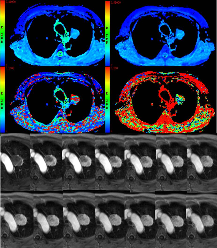
57 consecutive patients with solitary pulmonary nodules underwent DW-IVIM (multi-b-factor DWI with b values of 0, 20,50, 100, 150,200, 400, 600, and 1000 s/mm2) and DCE-3.0T MRI. ADCtotal, Tissue diffusivity (D), pseudo-diffusion coefficient (D*), and perfusion fraction (F) were calculated with mono--exponential and IVIM models. MER,Tmax, SLE and washout were calculated with semi-quantitative DCE analysis. Receiver operating characteristic (ROC) curves was constructed to estimate the diagnostic performance of both methods in differentiating diagnosis and the optimal cut-off values were obtained.Results
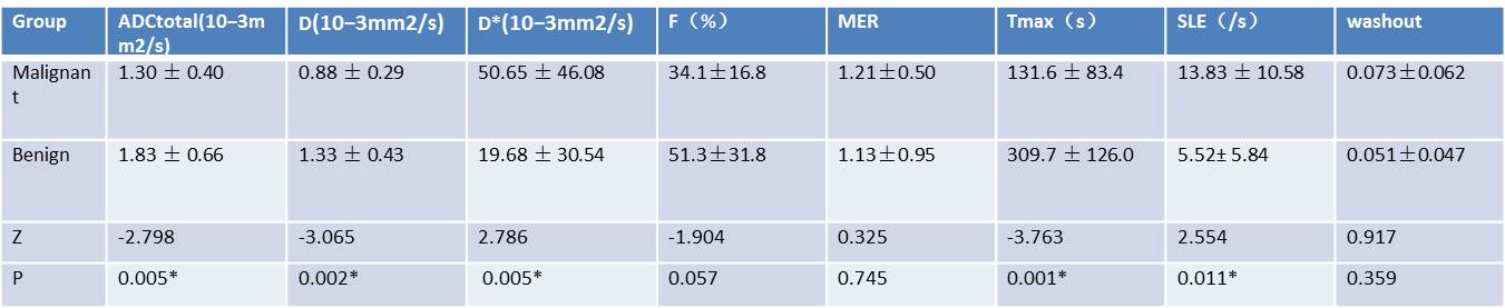
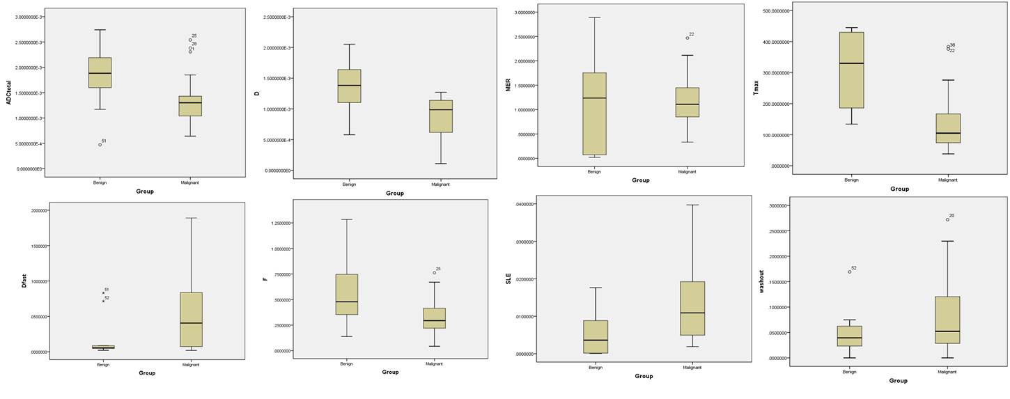
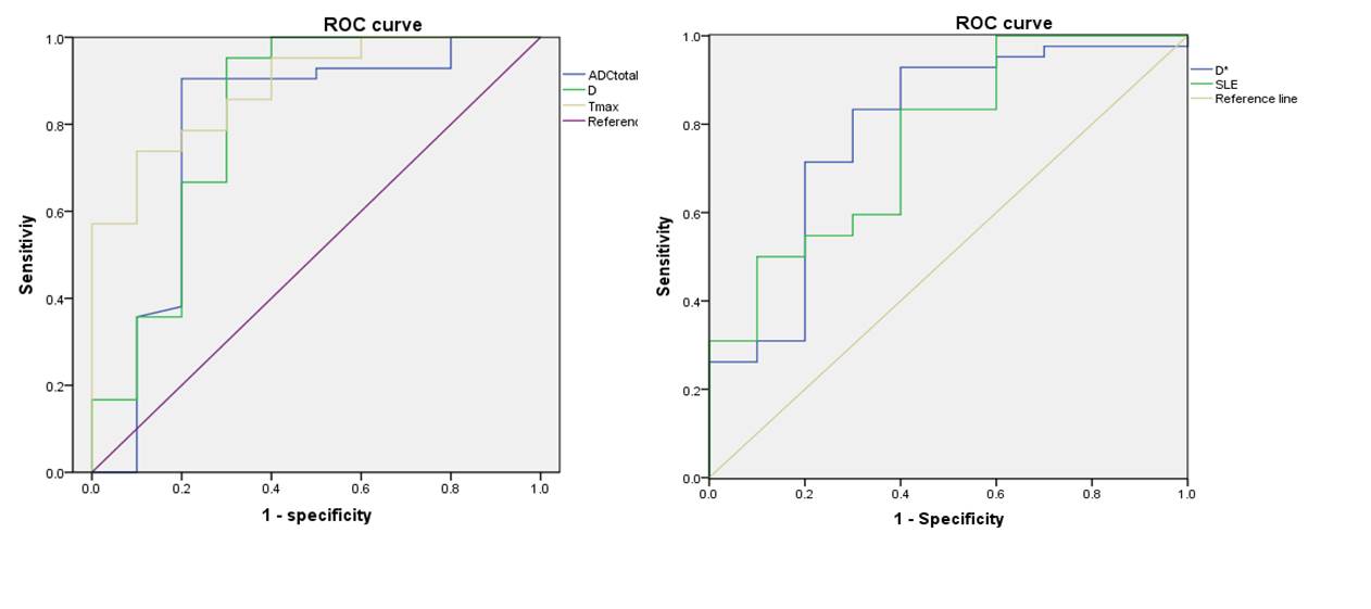
ADCtotal and D were significantly higher for benign lesions than for LC ([1.83 ± 0.66] × 10-3 mm2/s vs. [1.30 ± 0.40] × 10-3 mm2/s; P = 0.005; [1.33 ± 0.43] × 10-3 mm2/s vs. [0.88 ± 0.29] × 10-3 mm2/s; P = 0.002). D*was found to be significantly higher in LC than benignity ([50.65 ± 46.08] × 10-3 mm2/s vs. [19.68 ± 30.54] × 10-3 mm2/s; P = 0.005). No difference was observed in F between LC and benignity (P = 0.06).By DCE-MRI,Tmax were significantly shorter in LC than that of benignity([131.6 ± 83.4] s vs. [309.7 ± 126.0] s; P < 0.001),SLE was significantly higher for LC than for benignity([13.83 ± 10.58] × 10-3 /s vs. [5.52± 5.84] × 10-3 /s; P = 0.011). Among ADCtotal,D,D*,Tmax and SLE,D and Tmax had better diagnostic effecacy with higher sensitivity(95.2%,85.7%) and accuracy(86.6%,76.8%) than other indices. The diagnostic performance can be improved by combined D and Tmax, leading to a sensitivity, specificity and accuracy of 97.6%, 93.4%, and 92.5%, respectively. Poor correlations were found between parameters derived from IVIM and DCE-MRI.Discussion
Diffusion-related parameters ADCtotal and D were lower in malignancy due to microstructure change of tumor cells, D value show higher sensitivity and accuracy, indicating IVIM model better reflect true diffusion of tumor cell water molecules. D* with high variability indicate the instability and made the parameter of limited application. DCE-derived parameters Tmax and SLE were significant different in LC and benignity, Shorter Tmax and higher SLE reflect angiogenesis and structure destruction of tumor tissue. Poor correlations between parameters derived from IVIM and DCE-MRI may demonstrate different mechanism of different protocols but a combination of both protocols can get an excellent differential performance.Conclusion
Both IVIM model based on DWI and DCE-MRI were useful for discriminating benign lesions from malignant pulmonary nodules, and can provide quantitative diffusion and perfusion information by multiple parameters. A combined DCE and IVIM could provide a more promising way in determination microstructure characteristics of solitary pulmonary nodules.Acknowledgements
No acknowledgement found.References
[1].Deng Y, Li X, Lei Y, et al. Use of diffusion-weighted magnetic resonance imaging to distinguish between lung cancer and focal inflammatory lesions: a comparison of intravoxel incoherent motion derived parameters and apparent diffusion coefficient.[J]. Acta Radiologica, 2015.
[2].Wang L L, Lin J, Liu K, et al. Intravoxel incoherent motion diffusion-weighted MR imaging in differentiation of lung cancer from obstructive lung consolidation: comparison and correlation with pharmacokinetic analysis from dynamic contrast-enhanced MR imaging.[J]. European Radiology, 2014, 24(8):1914-22.
Figures