2058
The investigation of the relationship between obstetrical risk factors and pelvic floor injuries: a MRI-based studyLimei Guo1, Yujiao Zhao1, Zhizheng Zhuo1, and Wen Shen1
1Tianjin First Center Clinical College, Tianjin, People's Republic of China
Synopsis
Vaginal childbirth is an important cause to pelvic floor injuries. In this study, we aim to identify various forms of the injuries and the association with obstetrical risk factors in primiparous women. The results showed that there were significant differences about the proportions of various patterns of the pelvic floor injuries and the severity of LAM injuries between the groups with and without obstetrical risk factors.
Purpose
Vaginal childbirth is an important cause to levator ani muscle(LAM) avulsion. At present, the majority of study is about LAM injuries during childbirth based on ultrasound imaging. MRI has an advantage on the contrast of soft tissue and bone over ultrasound imaging, and has the ability to detect the lesions of pubic bones. In addition, there are few studies on the relationship between of obstetrical risk factors and the pelvic floor lesions. In this study, we aim to identify the forms of pelvic floor injuries and the association with obstetrical risk factors in primiparous women.Methods
A prospective study were carried out with 115 primiparous women after childbirth, and 34 of them have suffered obstetrical risk factors (such as instrumental delivery, childbirth weight≥4000g, the second stage of labor≥120min, precipitated labor, maternal age ≥35)1 and the remaining 81 women suffered no any risk. All the subjects underwent an MR examination of the pelvic floor six weeks after delivery by using axial and coronal T2WI FSE technique and axial T2WI FSE technique with fat saturation. All the MR examinations were carried out based on a 3T MR scanner (Ingenia, Philips Healthcare, the Netherlands). Two radiologists with more than 5 years experiences and blinded to obstetric and delivery factors reviewed the forms and severity of pelvic floor injuries. The injurie types were defined as follows (images shown in Figure 1 and Figure 2): (1) LAM tears which were evaluated for discontinuity of muscle fiber and observed as loss of visible muscle in an area. And this type injury was furtherly scored based on the lateral side score standard: score 0 with no muscle fiber deficiency, score 1 with less than 50% muscle fiber deficiency, score 2 with more than 50% muscle fiber deficiency, score 3 with completely muscle fiber loss, and if a sum score of bilateral sides was 0-3, then evaluated as minor injury and if sum score was 4-6, then evaluated as major; (2) pubic bone injuries which means there are only edema lesions or the lesions of fracture and edema. The types of injuries and risk factors were analyzed by using Chi-square test.Results
The statistical results are summarized in Table 1. There were significant differences about the proportions of various patterns of the pelvic floor injuries and the severity of LAM injuries between the groups with and without obstetrical risk factors.Discussion
LAM tears were more likely to occur in the women with risk factors. The cause is supposed to be that these factors can contribute to a more overstress of LAM during delivery. And there was no difference about the presence of the bone injuries in the two groups, which may indicate that the mechanism of the bone injuries differs from LAM. In the future, the relationship of some certain factors and LAM lesion need a further exploration. And a long-term follow-up of the pelvic floor injuries would be carried out to identify whether there is some correlation about these lesions and pelvic floor dysfunction.Conclusions:
There are some relationship between obstetrical risk factors and pelvic floor injuries. The obstetrical factors might predict increased risk of LAM injuries, but pubic bone injuries are independent of these factors.Acknowledgements
None.References
[1] Memon, H. U., & Handa, V. L. (2013). Vaginal childbirth and pelvic floor disorders. Womens Health (Lond), 9(3), 265-277; quiz 276-267. doi: 10.2217/whe.13.17Figures
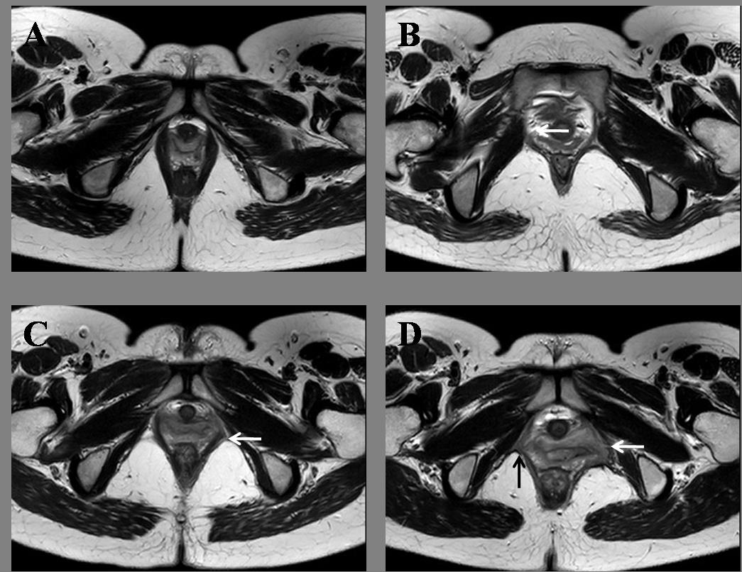
Figure 1. Different scores
of levator ani injuries severity in axial magnetic resonance images. A. No
injuries in LAM, which were scored 0; B. loss of right muscle fiber less than 50%,
which were further scored 1; C. loss of left muscle fiber more than 50%, which were
scored 2; D. Black arrows identify loss of right muscle fiber more than 50%,
and white arrows represent complete deficiency,
which were scored 3.
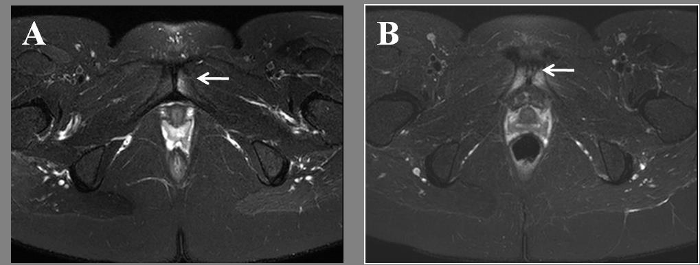
Figure 2. Example of forms of pubic bones
injuries in axial T2WI-FSE with fat saturation. A. left pubic bone edema; D. left pubic bone fracture
accompanied edema.
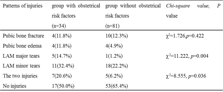
Table 1.
The patterns of pelvic floor injuries in the two groups and the statistical
results (Chi-square Test, p<0.05 indicate a significant difference)