2054
A new technique of SPIO-enhanced MRI: delayed recovery of T2*-weighted signal intensity as a novel diagnostic marker for visualization of irradiated liver parenchyma.1Division of Functional Imaging, Exploratory Oncology Research & Clinical Trial Center, National Cancer Center, Kashiwa, Japan, 2Department of Radiology, Graduate School of Medicine, University of Tokyo, Tokyo, Japan, 3Department of Radiology, Graduate School of Comprehensive Human Sciences, University of Tsukuba, Tsukuba, Japan
Synopsis
Visualization of irradiated liver parenchyma may assist safety margin assessment in radiotherapy. We demonstrate that an MR imaging technique has the ability to visualize irradiated liver parenchyma after 30-Gy irradiation in a tumor-bearing rat model. In this technique, superparamagnetic iron oxide (SPIO) is administered to label Kupffer cells (KCs) before, rather than after irradiation. A dose of 30-Gy is a lower, more clinically relevant dose than that used in the previous studies. Our results suggest that 30-Gy irradiation delays the recovery of hepatic T2*-weighted signal after SPIO administration. Presumably, irradiation delays degradation process of SPIO in the KCs.
Purpose
Radiotherapy is a less invasive option in selected patients who have liver cancer, although surgical resection is a current standard therapy. Stereotactic body irradiation therapy (SBRT) can deliver high-dose irradiation (approximately 30-Gy or more) to liver tumors while minimizing the radiation dose to adjacent liver parenchyma. Safety margin assessment may provide a reliable indicator of a risk of local recurrence after SBRT; however, such assessment is currently not possible during the treatment period or soon after its completion because a clear and simultaneous visualization of the irradiated liver parenchyma and liver cancer remains challenging with currently available imaging techniques. Recently, Furuta et al. have reported a new magnetic resonance imaging (MRI) technique that visualizes the irradiated liver parenchyma and liver cancer simultaneously by using gradient-echo T2*-weighted images (T2*WIs).1 They administered superparamagnetic iron oxide (SPIO) to label Kupffer cells (KCs) before, rather than after, 50 to 70-Gy irradiation to the tumor-bearing liver lobe of rats. They demonstrated that the irradiated liver parenchyma and liver tumor appeared as low and high signal intensities at 7th day after irradiation. Noticeably, the presence of the low signal intensity was associated with tumor regression. In the present study, we examined whether the same MRI technique has the ability to visualize irradiated liver parenchyma, non-irradiated liver parenchyma, and liver cancer even after 30-Gy irradiation, which is a lower, more clinically relevant dose than that used in the above-mentioned study.Methods
We inoculated N1-S1 hepatoma cells to the left liver lobe (LL) of 5 female Sprague-Dawley rats as described previously.2 One week later, we administered 20 μmol iron/kg body weight of SPIO (Resovist; Bayer Yakuhin, Ltd., Japan) to the rats and acquired respiratory-gated T2*WIs and T2WIs of the liver by using a 9.4-tesla scanner (BrukerBiospin, Ettlingen, Germany). We estimated tumor volumes on T2*WIs by using the ellipsoid formula. Four hours later, we delivered a single dose of 30-Gy irradiation to the tumor-bearing LL. On day 7, we examined the liver by using the same MRI technique without additional SPIO. We also performed T2* mapping of the liver by using Multiple Gradient Echo sequence (BrukerBiospin, TR/TE/FA = 1000/3.1-18.5 ms [5 steps]/25°). Relative signal intensity (RI) was calculated by dividing the signal intensities of the LL, right liver lobe (RL), and tumor by the signal intensity of the paraspinal muscle. We compared the differences in RIs of the LLs and RLs on days 0 and 7 by using Tukey’s test. We also compared the differences in T2* values of the LLs, RLs and paraspinal muscle on day 7 by using Tukey's test.Results
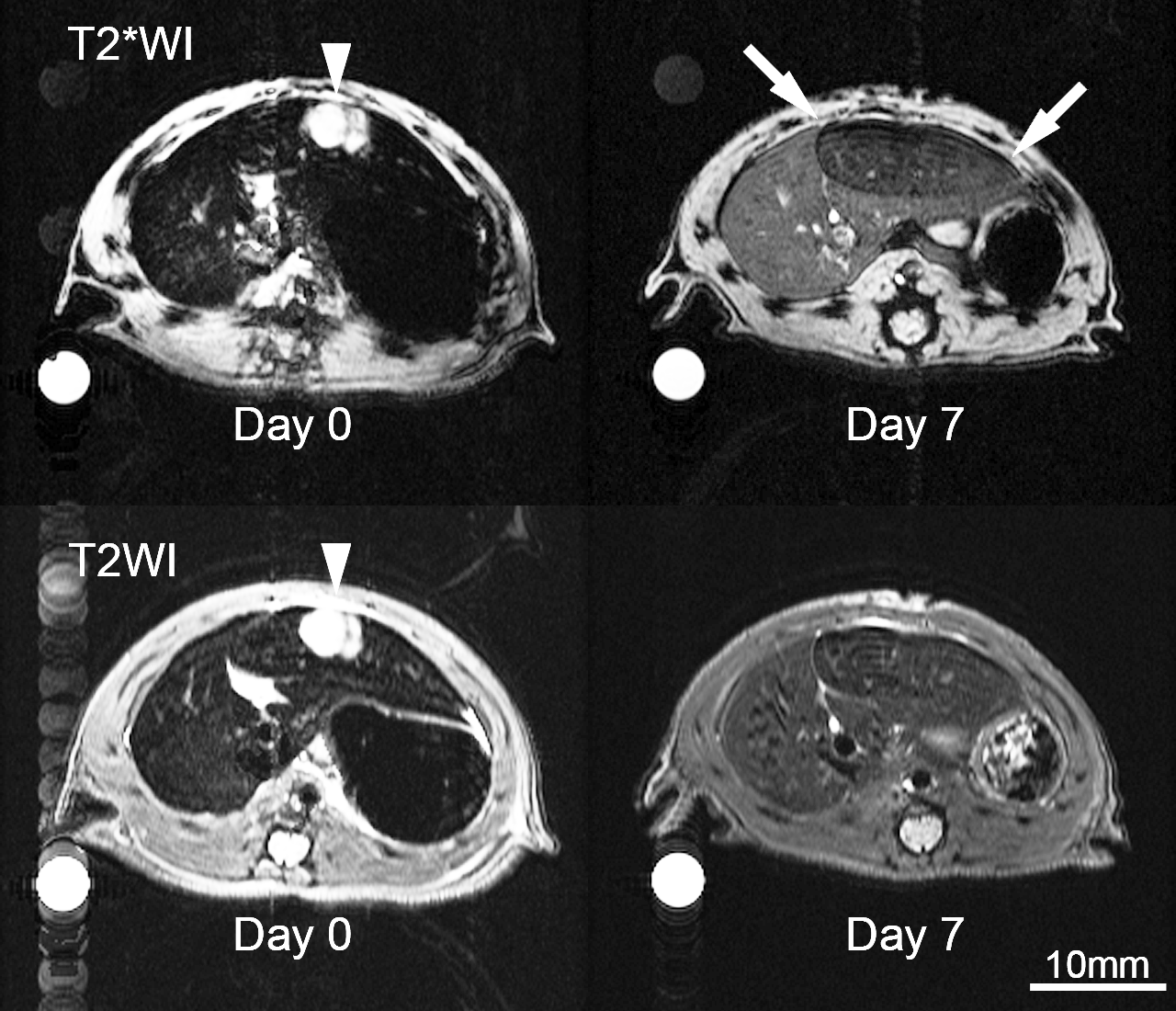
On day 0, tumor volumes were 166±27 mm3 (mean±standard error, n=5). RIs of the LLs and RLs were equivalently low in both T2*WIs (0.17±0.017, LL; 0.17±0.014, RL) and T2WIs (0.33±0.0067, LL; 0.29±0.0093, RL) due to SPIO accumulation. On day 7, we found no tumor in the liver in all rats. In the T2*WIs, RI of the LL (0.23±0.03) was significantly lower than that of the RL (0.52±0.03, p<0.001). In contrast, RI of the LL (0.46±0.06) and RL (0.56±0.06) did not differ significantly in T2WIs. T2* values of the LL (3.2±0.34 ms) were significantly lower than those of the RL (9.1±0.17 ms, p<0.05).Discussion
We found that T2*WI, not T2WI, has the ability to visualize irradiated liver parenchyma as low signal intensity areas at 7th day after 30-Gy irradiation to the previously SPIO-accumulated liver. In contrast, non-irradiated liver parenchyma regained the signal intensity during this period as KCs degraded SPIO particles to non-superparamagnetic iron ions.3 Thus, these findings suggested that 30-Gy irradiation delays the recovery of T2*-weighted signal intensity of the liver after SPIO administration. Presumably, irradiation delays degradation process of SPIO in the KCs as reported in the above-mentioned study.1 Lower T2* values of the irradiated liver parenchyma than those of non-irradiated liver parenchyma we observed supported this scenario because SPIOs produce magnetic susceptibility effect, thereby shortening T2* values of the liver. Since a single dose of 30-Gy irradiation resulted in complete regression of the tumors, we did not visualize safety margin (i.e., the irradiated liver parenchyma) surrounding the liver cancer lesion. We attributed the difference in tumor response between this study and the above-mentioned study1 to the difference in tumor volume at the time of irradiation. We treated smaller liver tumors; therefore, we observed complete response even by using smaller dose of irradiation.Conclusion
T2*WI has the ability to visualize irradiated liver parenchyma after 30-Gy irradiation to the previously SPIO-accumulated liver. The visualization of the irradiated liver parenchyma may assist safety margin assessment in radiotherapy in the clinic.Acknowledgements
This work was supported by JSPS KAKENHI Grant Number JP16K10332, Foundation for Promotion of Cancer Research, and Research grant from Japan Radiological Society supported by Bayer.
The authors thank Dr. Shusei Hamamichi for English proofreading.
References
1. Furuta T, Yamaguchi M, Minami M, et al. Persistent T2*-hypointensity of the liver parenchyma after irradiation to the SPIO-accumulated liver: An imaging marker for responses to radiotherapy in hepatic malignancies. J Magn Reson Imaging. 2016 Jul 4. Epub ahead of print
2. Yamaguchi M, Mitsuda M, Ezawa K, et al. Artifact-reduced simultaneous MRI of multiple rats with liver cancer using PROPELLER. J Magn Reson Imaging 2013;38(1):225–230.
3. Weissleder R, Stark DD, Engelstad BL, et al. Superparamagnetic iron oxide: pharmacokinetics and toxicity. AJR Am J Roentgenol. 1989;152:167–173.
Figures