2053
Parameter Optimization for Non-contrast-enhanced Renal MR Angiography and Its Age-dependent Preliminary Study1the First Hospital of China Medical University, Shenyang, People's Republic of China
Synopsis
Our purpose was to investigate the feasibility of NCE-MRA using Time-SLIP technique on healthy volunteers by determining the optimized TI value and its preliminary relationship with age as well. 61 healthy volunteers were recruited and divided into two age groups. The acquired data using six differernt TIs sequences were measured and analyzed to get three parameters including VKR, grade of renal artery branches and grade of imaging quality. In conclusion, the Time-SLIP technique is able to obtain renal MR angiography with optimized TI value 1500ms. Moreover, the age of individual subjects can affect the optimized TI value.
PURPOSE
With the incidence of renal artery stenosis ( RAS ) increased, the potential occurrence of contrast-induced nephropathy ( CIN ) and nephrogenic systemic fibrosis ( NSF ) may restrict the uses of CTA and MRA1,2, time spatial labeling inversion pulse ( Time-SLIP ) technique is one of the methods of non-contrast-enhanced renal MR angiography ( NCE-MRA ). Our purpose was to investigate the feasibility of NCE-MRA using Time-SLIP technique on healthy volunteers by determining the optimized inversion time ( TI ) value and discussing its preliminary relationship with age as well.METHODS
61 healthy volunteers ( range, 22-80 years ) were recruited in this study and divided into two groups ( >50 years, ≦50 years ). All the volunteers underwent 3.0T MRI ( Vantage Titan 3T, Toshiba, Japan ) examination with six differernt TIs sequences ( TIs=1200ms, 1300ms, 1400ms, 1500ms, 1600ms, 1700ms ) . The acquired data were measured and analyzed to get three parameters respectively, including vessel-to-kidney ratio ( VKR )3, grade of renal artery branches , grade of renal artery imaging quality.RESULTS
One example of MIP images was shown in figure1. The VKR was highest when the TI value was 1500ms both in the left and right kidneys ( figure2 ). The scores of the renal arteries branches were gradually rising up but tending to be steady higher than 1400ms ( figure3 ). The scores of renal artery image were the highest at TI = 1500ms/1600ms in all subjects ( figure4 ). In subjects above 50 years old, the optimized TI value was 1600ms, but the highest score of imaging quality is in subjects with TI=1500ms below 50 years old ( 50 years included ). Moreover, every setion of renal arteries in subjects below 50 years old ( 50 years included ) acquired the higher score of imaging quality comparing to another group. The imaging quality of all sections of renal arteries of all subjects and two groups was shown in table 1.DISCUSSION
There are several factors that affect the optimized TI value, including imaging signal intensity, the display condition of renal artery branches , the imaging quality of renal arteries and so on. In different TI sequences, the VKR was the highest at TI=1500ms group, but there is not statistical significance between 1500ms and 1600ms. At TI=1200ms-1500ms, the VKR values were increasing gradually with the extension of TI time, which indicates that the inflow of renal blood from the abdominal aorta to the branches of renal arteries resulting in the renal trunk full with blood. However, with the inversion signal of renal parenchyma and the background have been recovered, the image quality of renal artery became worse which brought about that the VKR values were tending to decrease with the TI continuous increased4. The branches of renal became better along with the prolong of TI time, but is tending to be steady after 1400ms. This is because that three sections of renal arteries have been full of blood eventually. The position of TI pulse and respiratory of subjects can all affect the imaging quality of renal arteries, so coronal scanning with the TI pulse parallel to the upper limb of kidneys was required in this study5-7. Furthermore, all subjects were told to breathe steadily during Time-SLIP scanning. The subjects’ age is another factor that affect the optimized TI value. In the group above 50 years old, the score of imaging quality was the highest with the optimized TI value 1600ms, whereas the highest score of image quality was acquired in the group below 50 years old ( 50 years included ) with optimized TI=1500ms. Moreover, every setion of renal arteries in subjects below 50 years old ( 50 years included ) had the higher score of imaging quality comparing to another groups. These results may result from the hemorheological changes with different ages8,9.CONCLUSION
The Time-SLIP technique is able to obtain renal MR angiography successfully without the use of contrast medium, whose optimized TI value is 1500ms. Moreover, the subjects’ age is one of the factors that affect the optimized TI value.Acknowledgements
No acknowledgement found.References
1. Kume K, Yasuoka Y, Adachi H, et al. Impact of contrast-induced acute kidney injury on outcomes in patients with ST-segment elevation myocardial infarction undergoing primary percutaneous coronary intervention ?. 2013, 14(5):253-257.
2. Weller A, Barber J L, Øystein E Olsen. Gadolinium and nephrogenic systemic fibrosis: An update. Pediatric Nephrology, 2013, 29(10):1927-37.
3. Kurata Y, Kido A, Fujimoto K, et al. Optimization of non-contrast-enhanced MR angiography of the renal artery with three-dimensional balanced steady-state free-precession and time-spatial labeling inversion pulse (time-SLIP) at 3T MRI, in relation to age and blood velocity. Abdominal Radiology, 2016, 41(1):1-8.
4. Brucher N, Vial J, Baunin C, et al. Non-contrast-enhanced MR angiography using time-spin labelling inversion pulse technique for detecting crossing renal vessels in children with symptomatic ureteropelvic junction obstruction: comparison with surgical findings. European Radiology, 2016,26(8):2697-704.
5. Zhang Y, Xing Z, Liu Y, et al. Nonenhanced renal MR angiography using steady-state free precession (SSFP) and time-spatial labeling inversion pulse (Time-SLIP): repeatability and comparison of different tagging location. Abdominal Imaging, 2014, 39(5):1000-8.
6. Liu X, Berg N, Sheehan J, et al. Renal transplant: nonenhanced renal MR angiography with magnetization-prepared steady-state free precession. Radiology, 2009, 251(2):535-42.
7. Albert T S, Akahane M, Parienty I, et al. An International Multicenter Comparison of Time-SLIP Unenhanced MR Angiography and Contrast-Enhanced CT Angiography for Assessing Renal Artery Stenosis: The Renal Artery Contrast-Free Trial. Ajr American Journal of Roentgenology, 2015, 204(1):182-8.
8. Ting-yu H , Wei-ying Z,et al. Specificity of Hemorrheological Test in Different Age Diseases. Chinese Journal of Hemorheology, 2004, 14(1):68-71.
9. Mowat D H R, Haites N E, Rawles J M. Aortic blood velocity measurement in healthy adults using a simple ultrasound technique. Cardiovascular Research, 1983, 17(2):75-80.
Figures
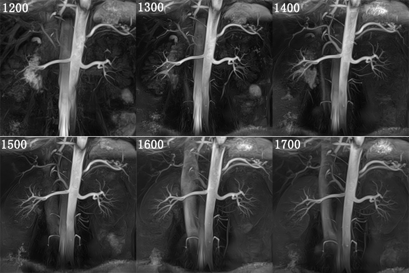
Figure1 MIP images of a 35 years old man
The branches of renal arteries were shown better and better along with the prolong of TI time, but the distal branches became saturated after 1400ms; the image quality of renal trunk and all the branches became better, which were the best when the TI was close to 1500ms. However, with the continous prolong of TI time, the recovered background signal made the image quality of renal arteries worse.
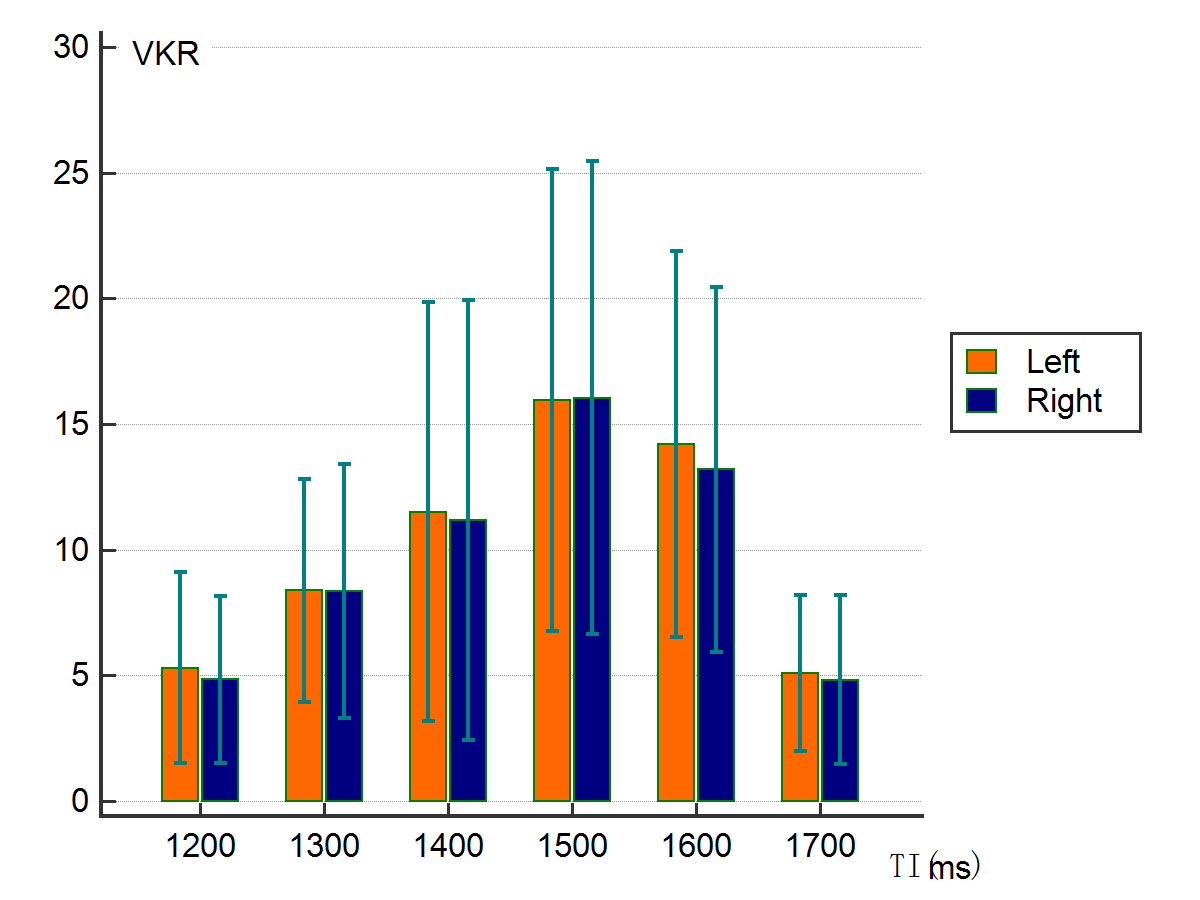
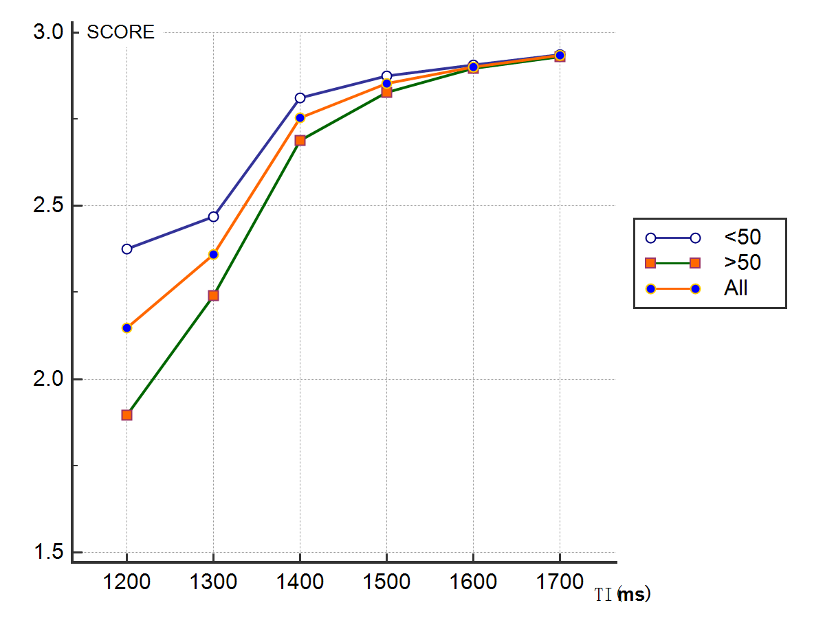
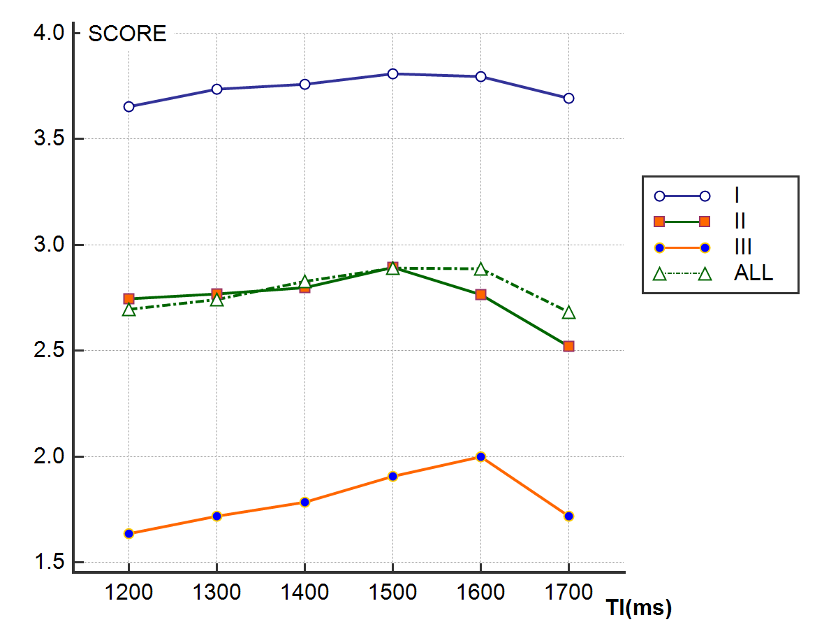
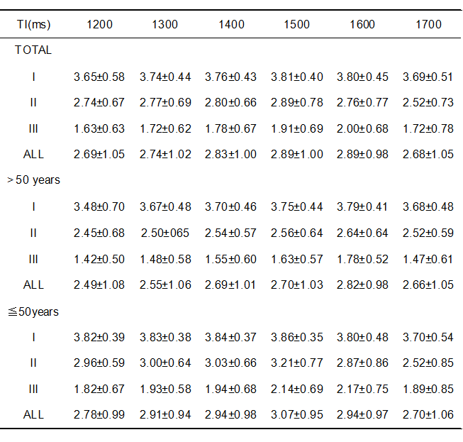
Table1 Imaging quality grade of renal arteries in all subjects and two age groups
I:section I, renal trunk; II: section II, renal arteries in renal hilus; III: section III, renal arteries in renal parenchyma; ALL: all sections