2051
Cyclic Changes of the Boundary Sharpness of Uterine Zonal Structures Visualized by High-resolution T2-weighted Images in Young and Middle-aged Females during the Menstrual Cycle1Radiology, Peking Union Medical College Hospital, Beijing, People's Republic of China, 22. MR Collaboration NE Asia, Siemens Healthcare, Beijing, People's Republic of China, 3OB&GYN, Peking Union Medical College Hospital, Beijing, People's Republic of China
Synopsis
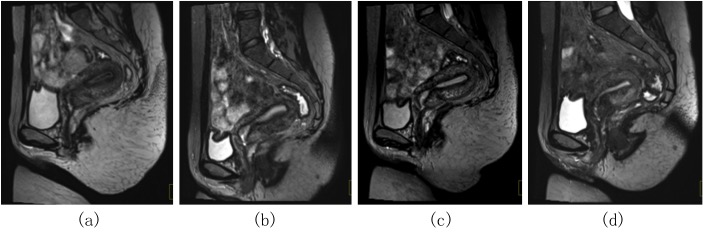
This study aimed to demonstrate the cyclic changes of the boundary sharpness of uterine three zonal structures of young and middle-aged women on 3T using a 3D T2-weighted SPACE sequence during the menstrual cycle. Peri-ovulatory phase exhibited the clearest boundary sharpness of corpus zonal structures following by FP, LP and MP, while that of the cervix were almost well-defined during the menstrual cycle.
Introduction/Purpose
The uterus was visualized as three zonal structures on T2-weighted images. Assessment of the boundary sharpness and consecutiveness of these zonal structures plays an important role in staging evaluation of uterine neoplasm. Previous studies found that some manifestations of the uterus showed cyclic changes during the menstrual cycle. The purpose of this study was to investigate cyclic changes of the boundary sharpness of the three zonal structures of uterine corpus and cervix on high-resolution 3D T2-weighted MR images in different age groups during the menstrual cycle.Methods and materials
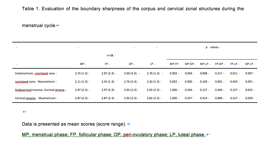
38 normal volunteers (20-40yrs, mean 29yrs)(20-30yrs, n=22; 31-40yrs, n=16) with regular menstrual cycle accepted repeated pelvic MR examinations on a MAGNETOM Skyra 3T MR scanner (Siemens, Erlangen, Germany) on the 2nd or 3rd day of menstrual phase (MP), follicular phase (FP), peri-ovulatory phase (OP) and luteal phase (LP). The 3D high-resolution T2-weighted images were acquired using a SPACE sequence with the following parameters: TR/TE = 1500/136 ms, FOV = 300 × 300 mm2, slice thickness = 0.9 mm, voxel size = 0.9 × 0.9 × 0.9 mm3, total acquisition time = 7:59 min. Two radiologists blinded to the clinical data and scanning phase evaluated the boundary sharpness of the three zonal structures of uterine corpus and cervix on mid-sagittal images on a 3-point Likert-scale (3, well-defined; 2, obscure; 1,ill-defined) independently. T2 signal intensity of each zonal structure was measured, and SI ratio between adjacent structures was calculated. Inter-observer agreement was evaluated by Cohen’s Kappa analysis. Paired Wilcoxon and t-test were used to evaluate the differences between age groups during the four phases.Results
The boundary sharpness of the uterine three zonal structures showed no significant difference between the two age groups (p>0.05). Only 10.5% (4/38) of volunteers, all in the 20-30 yrs group, exhibited no variation during the menstrual cycle, which were all well-defined. Mean scores of boundary sharpness of the corpus zonal structures from high to low were OP, FP, LP, MP with significant differences (Table 1), corresponding to the SI ratio results. The boundary sharpness of the cervical zonal structures showed no statistical difference during the menstrual cycle (p>0.05) (Table 1), which exhibited almost well-defined. Inter-observer agreement was excellent (all kappa >0.75).
Discussion
The present study used 3D high-resolution T2-weighted imaging to enable interactive multiplanar reformatting at arbitrary orientations and to evaluate the cyclic changes of the boundary sharpness of the uterine zonal structures during the menstrual cycle. The peri-ovulatory phase could be selected as the best phase to investigate zonal related uterine disease, especially in the evaluation of the depth of myometrium invasion of some gynecological malignancies. However, the phase of menstrual cycle has little influence on the depiction of cervical zonal structures.Conclusion
Boundary sharpness of three zonal structures of the uterine corpus showed dynamic variation during the menstrual cycle, and the peri-ovulatory phase exhibited the clearest boundaries. Cervical zonal structures were almost well-defined throughout the menstrual cycle.Acknowledgements
None.References
1. Hori M, Kim T, Onishi H, et al. Endometrial cancer: preoperative staging using three-dimensional T2-weighted turbo spin-echo and diffusion-weighted MRI at 3.0 T: a prospective comparative study. Eur Radiol 2013;23:2296-305.
2. Hori M, Kim T, Onishi H, et al. Uterine tumors: comparison of 3D versus 2D T2-weighted turbo spin-echo MRI at 3.0 T - initial experience. Radiology 2011;258:154-63.
3. He YL, Ding N, Li Y, et al. Cyclic changes of the junctional zone on 3 T MRI images in young and middle-aged females during the menstrual cycle. Clin Radiol. 2016;71:341-8.
Figures