2045
MRI to evaluate the response of the locally advanced cervical cancer to CCRT: MRS(magnetic resonance spectroscopy), DWI(diffusion weighted image), and T2WIByung Chul Kang1 and Hye Ran Hyun2
1Radiology, Mokdong Hsopital, EWUMC, Seoul, Korea, Republic of, 2Radiology, Mokdong Hospital, EWUMC, Seoul, Korea, Republic of
Synopsis
MRS(MR Spectroscopy) as well as T2Wi, and diffusion weighted images may be used to evaluate the response of LACC to CCRT.
INTRODUCTION & OBJECTIVE:
The objective of this study
was to investigate whether T2 weighted images, MR spectroscopy, diffusion-weighted
imaging (DWI) of the locally advanced cervical cancer (LACC) could be used to
assess to post-treatment response of CCRT at the higher magnetic field strength
of 3.0 T.
METHODS:
Retrospectively thirty nine
women with LACC received magnetic resonance were enrolled at one more different
times after CCRT. If high signal intensity was still observed on T2WI after
CCRT we considered as viable cancerous tumor area. In MRS pattern, we
considered as positive viable cancer if the calculated choline peak were 2
times than the creatinine peak. In DWI, we considered as positive viable if
high signal intensity observed in cervix. Confirmative diagnoses were
determined with PET-CT or cervical biopsy. Accuracy rates were analyzed between
MR images and confirmative diagnosis statistically. For the statistical
analysis, chi-square test was used with SPSS version 18.0.
RESULTS:
73 MR studies of 39 patients
were obtained after CCRT. Diagnostic accuracy rates were 66% in T2Wi, 73.1% in
Diffusion-weighted images, and 66.7% in MRS. Correlations of MRS and DWI with
the cervical biopsy or PET-CT were all statistically significant (p= 0.01 in
MRS, p=0.008 in DWI). Correlations of T2Wi with the cervical biopsy or PET-CT
were not statistically significant (p= 0.02).
CONCLUSIONS:
MRS as well as DWI may be used
to evaluate the response of LACC to CCRT.
Acknowledgements
We appreciate the hospital collegues for helping to acquire the excellent MR spectroscopic datas.References
Recurrent uterine cancer afte surgery : MRI pattern and their changes after concomitant chemoradiation. Radiol Med 2008;113:1143-1156
Evaluation PET-CT in the detection and management of recurrent cervical cancer: systematic review of diagnsotic accuracy and sugjective elicitation. Royal College of Obstetrician and Gynecologists 2013;10.1111/1471-0528.12488
Figures
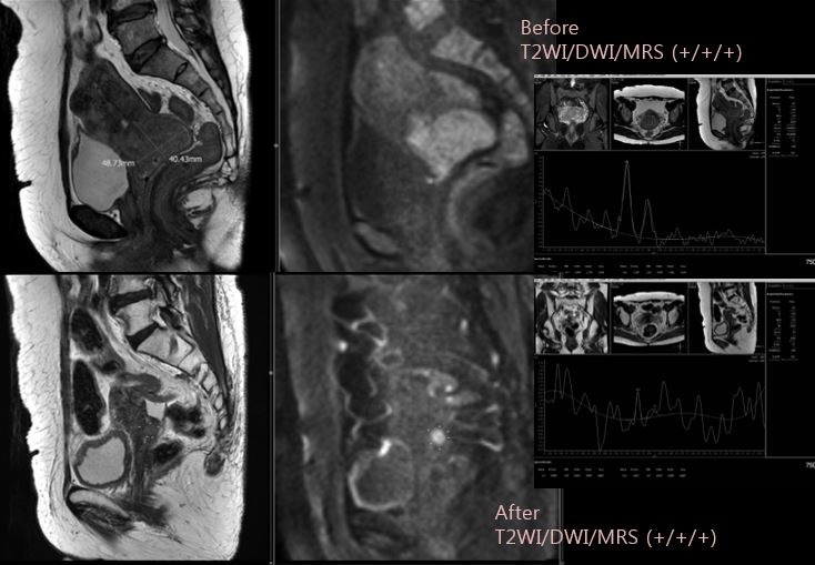
Before CCRT, signals of the cervical cancer show positive on T2WI, DWI, and MRS(MR spectroscopy). Even after CCRT, signals of the cervix cancer shows also positive on T2WI, DWI, and MRS.
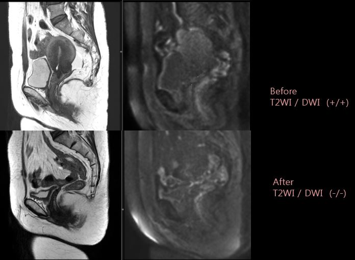
Befroe CCRT, signals of the cervical cancer show positive on T2WI, and DWI. After CCRT, signals of the cervix cancer shows negative on T2WI, and DWI.