2039
Computed Diffusion-Weighted Image for Abdominal MRI1Advanced Biomedical Imaging Research Center, Kobe University Graduate School of Medicine, Kobe, Japan, 2Center of Radiology and Radiation Oncology, Kobe University Hospital, Kobe, Japan, 3Toshiba Medical Systems Corporation, Otawara, Japan, 4Radiology, Kobe University Graduate School of Medicine, Kobe, Japan
Synopsis
The purpose of this study was to assess capability of computed DWI in evaluation of various abdominal diseases. We found cDWI can improve image quality and malignant lesion contrast, conspicuity, and detection. cDWI is a useful post-processing tool for abdominal MRI.
BACKGROUND & PURPOSE
Remaining problems in abdominal DWI
•Contamination of perfusion effect at lower b values
•Low SNR at higher b values
•Inadequate signal suppression of fluid
•Image distortions due to respiration or intestinal air
•Difficulties in preset of optimal b value
•Expertise is required for interpretation of ADC map
Computed DWI (cDWI) software can reconstruct DWI at any b value as post-processing.
The purpose of this study was to assess cDWI in evaluation of abdominal diseases.
MATERIALS & METHODS
Patients & Abdominal Lesions
•102 patients (52 men and 50 women, mean: 67.3 years), who were suspected to have hepato-biliary-pancreatic malignancy and underwent 3T-MRI, were retrospectively analyzed.
•65 malignant (15 HCCs, 4 CCCs, 11 metastatic liver, 9 bile duct, 5 gallbladder, 17 pancreatic CAs, etc.) and 68 benign (21 renal cysts, 14 hepatic cysts, 7 hepatic hemangiomas, 6 bilomas, 2 liver abscesses, 6 IPMNs, etc.) lesions were confirmed and chosen for analysis.
Imaging Technique
•All patients underwent MRI at a 3T scanner (Vantage Titan 3T; Toshiba Medical Systems Co., Otawara, Japan).
•Actual DWIs were obtained with SE-EPI sequence (TR/TE/FA=6500-11100/70/90, b values: 0, 1000, matrix: 128×128, thk: 4-7 mm, 26-40 slice, Nex: 4, scan time: 4-6 min, PASTA+SPAIR, PI: 2.2 (y), MPG: y, z) as one of routine sequences in our institution.
cDWI Reconstruction
• cDWI images at b values of 200, 400, 600, 800, 1200, 1400, 1600, 1800, and 2000 were reconstructed as DICOM-formatted images using a personal computer and prototype software (Toshiba Medical Systems Co.).
Image Analysis
Quantitative
•Signal intensities (SI) in the organs and lesions were measured.
•SIs of each organ were compared among the images.
•SNRs of each organ (SNR=SIorgan/SDorgan) were compared among the images.
•Lesion contrasts (CM = SIlesion-SIorgan / SIlesion+SIorgan) were measured and compared among the images.
Qualitative
•Two readers recorded b values with best quality and with complete gallbladder signal suppression, and assessed lesion conspicuity on DWI1000 and cDWIs.
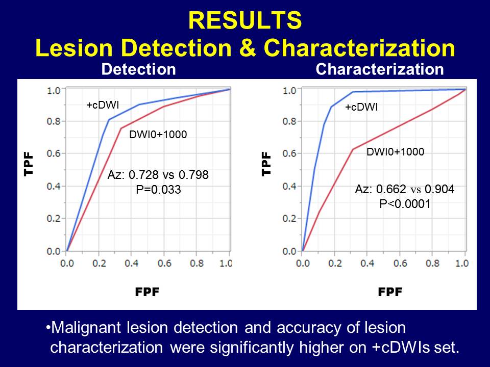
•Malignant lesion detection and accuracy of lesion characterization were assessed on DWI0+1000 and +cDWIs sets and compared using ROC analysis.
RESULTS
Quantitative
•SNRs were significantly highest on cDWI800 in the liver and on cDWI600 in the pancreas and spleen (P<0.0001).
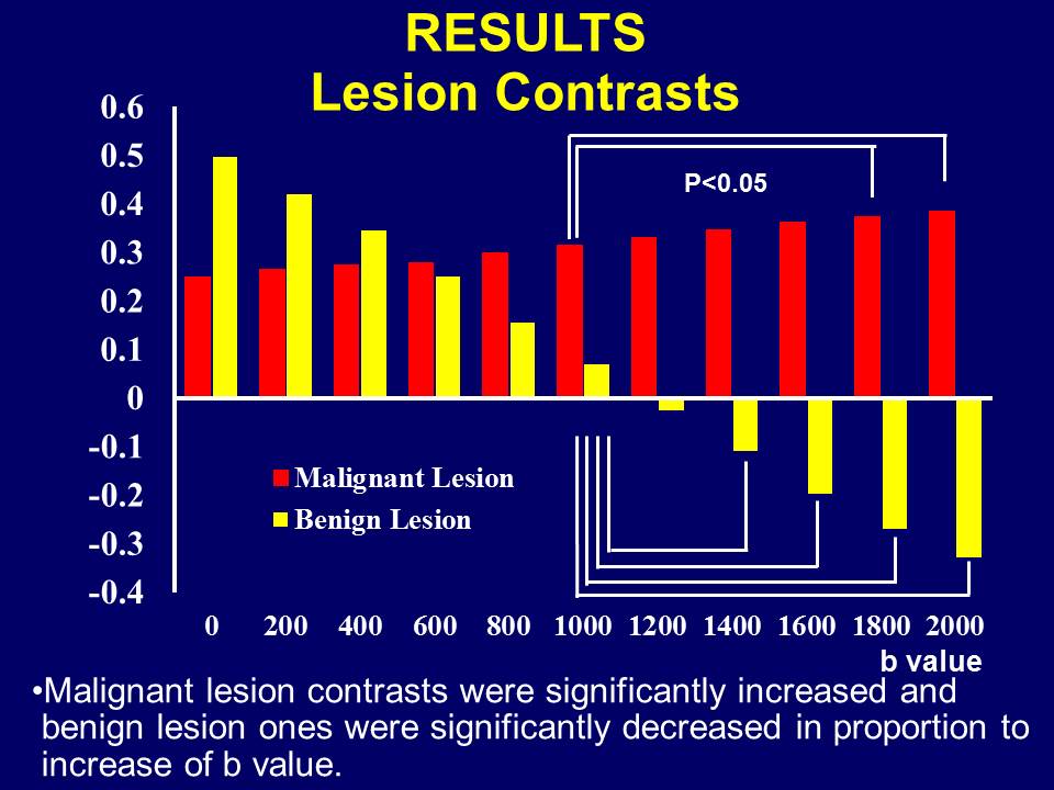
•Malignant lesions contrasts were significantly increased (0.026) and benign ones were significantly decreased (<0.0001) in proportion to increase of b value.
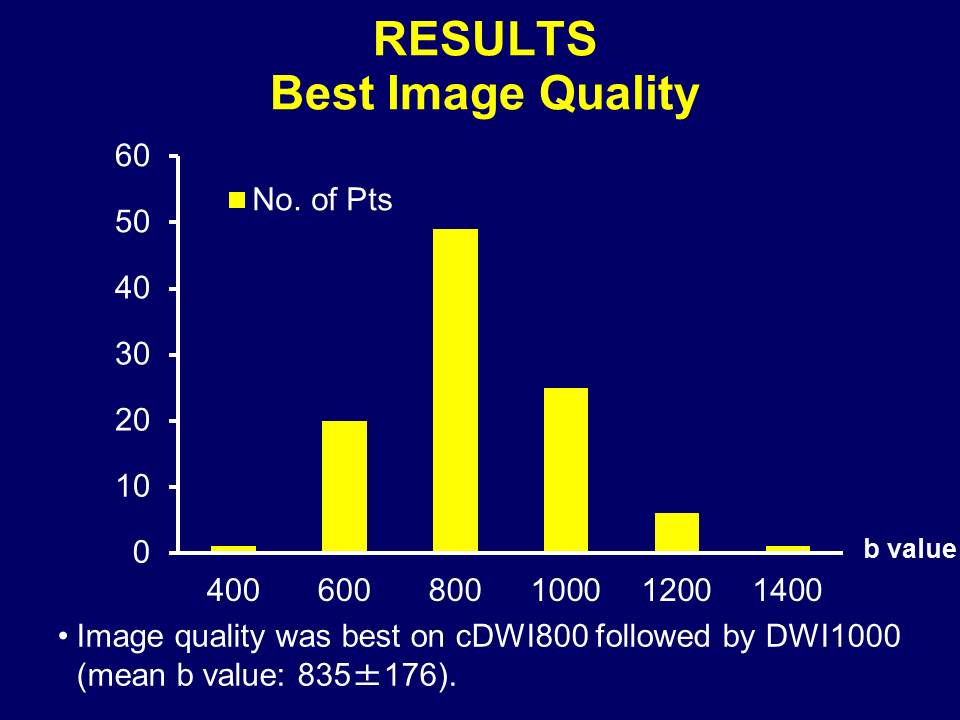
•Image quality was best on cDWI800 followed by DWI1000 (mean b: 835±176).
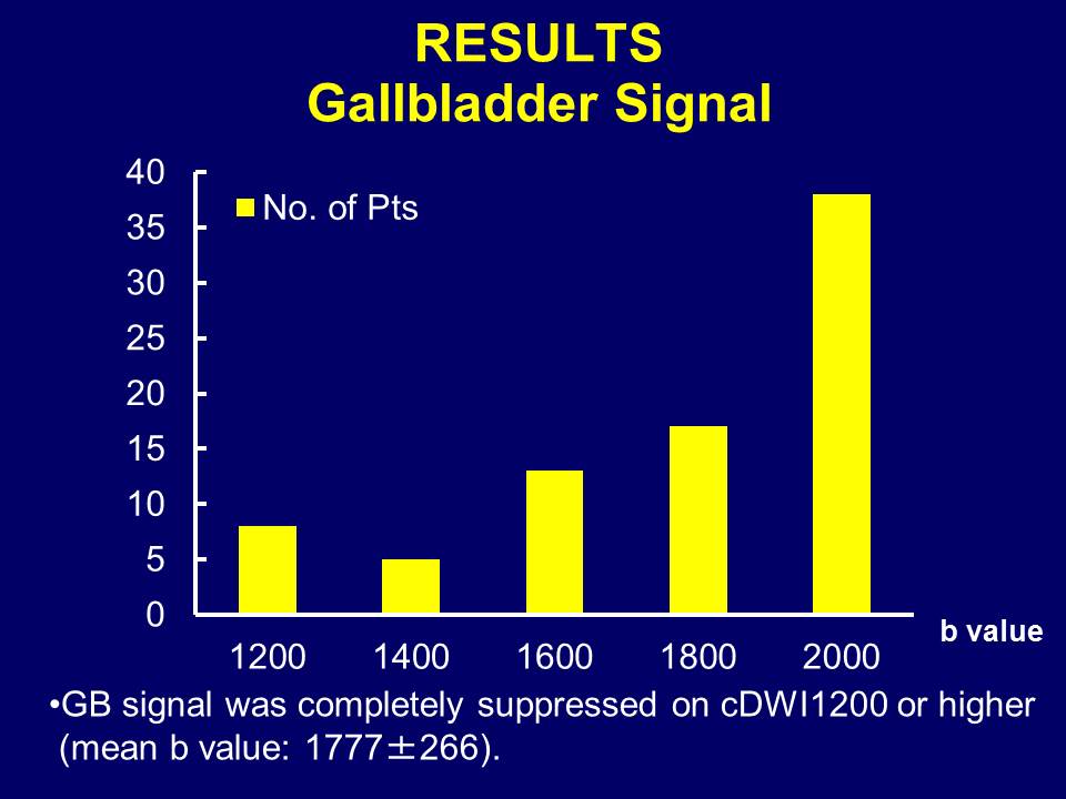
•GB signal was completely suppressed on cDWI1200 or higher (mean b: 1777±266).
Qualitative
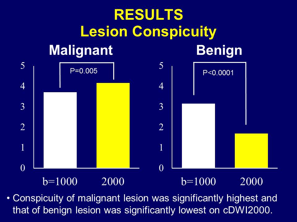
•Conspicuity of malignant lesion was significantly highest (0.005) and that of benign lesion was significantly lowest (<0.0001) on cDWI2000.
•Malignant lesion detection was significantly higher (Az: 0.728 vs 0.798, 0.033) and accuracy of lesion characterization was significantly higher (Az: 0.662 vs 0.904, <0.0001) on +cDWIs set.
DISCUSSION
•cDWI software enables reconstruction of DW image with any b value after MR examinations with short time even on personal computers.
•It has the potential to improve image quality when used for additional image reconstructions at lower b values.
•When used for additional image reconstructions at higher b values, it can improve malignant lesion conspicuity and add useful information for diagnosis.
•This technique is also useful for estimating optimal b values in each organs and lesions.
•Perfusion effect cannot be eliminated when actual source DW images are acquired at too low b values.
•It should be noted that misregistrations on images can be observed.
CONCLUSION
Computed DWI is a useful post-processing tool for abdominal MRI.Acknowledgements
No acknowledgement found.References
Liver tumor
•Nakamura Y, et al. EJR open 2016.
•Kawahara S, et al. Clin Imaging 2016.
•Shimizu H, et al. Eur J Radiol 2013.
DWIBS/whole Body
•Blackledge MD, et al. Radiology 2011.
Prostate & Ovary
•10 papers
Figures