2037
Intravoxel incoherent motion (IVIM) and diffusion kurtosis imaging (DKI) for differential diagnosing hepatocellular carcinoma (HCC) from hepatic hemangioma (HHA).1The First Affiliated Hospital of Dalian Medical University, Dalian, People's Republic of China, 2MR Research, GE Healthcare, Beijing, People's Republic of China
Synopsis
Intravoxel incoherent motion (IVIM) imaging is an extension of diffusion weighted imaging (DWI) that can be used to investigate both diffusion and perfusion changes in tissues. DKI adapts a kurtosis based model to depict the non-Gaussian diffusion process, which could be caused by the presence of different barriers in cellular complex structures (e.g. cell membranes and organelle compartments).Initial application of DKI focused on neuroimaging, Recently, it has also been reported that DKI may help to assess response to treatment in HCC. Comparing the IVIM and DKI parameters between carcinoma (HCC) and hepatic hemangioma(HHA) we found that IVIM and DKI can supply many meritorious parameters, combining with the IVIM and DKI may help in increasing the sensitivity and specificity of antidiastole.
Synopsis
Intravoxel incoherent motion (IVIM) imaging is an extension of diffusion weighted imaging (DWI) that can be used to investigate both diffusion and perfusion changes in tissues. DKI adapts a kurtosis based model to depict the non-Gaussian diffusion process, which could be caused by the presence of different barriers in cellular complex structures (e.g. cell membranes and organelle compartments).Initial application of DKI focused on neuroimaging, Recently, it has also been reported that DKI may help to assess response to treatment in HCC[1]. Comparing the IVIM and DKI parameters between carcinoma (HCC) and hepatic hemangioma(HHA) we found that IVIM and DKI can supply many meritorious parameters, combining with the IVIM and DKI may help in increasing the sensitivity and specificity of antidiastole.Target audience
Physicians and scientists who are interested in the diagnosis of hepatocellular carcinoma (HCC) and hepatic hemangioma (HHA), as while as the application of IVIM and DKI in abdomen.Purpose
MRI is widely used for the evaluation of patients with liver diseases because of its excellence in the depiction of soft tissue by means of various physiologic and functional imaging sequences. In particular, Intravoxel incoherent motion (IVIM) and diffusion kurtosis imaging (DKI) been adopted to neuroimaging, prostate and soft tissue tumors.[2-5]Our study was to assess the IVIM and DKI parameters for differential diagnosing hepatocellular carcinoma (HCC) and hepatic hemangioma (HHA).Methods
Thirty-eight consecutive patients (25 men: 13 women, mean age, 59 years) with 21 HCCs, 17 HHAs were enrolled in this study and inspected conventional MR and IVIM-MR and DKI-MR examination with 1.5-T MR imager from January 2015 to October 2015. MRI was performed using a 1.5-T MR imager (GE-Signa HDXT) in a protocol containing the routine T1WI, T2WI, IVIM (b= 0, 10, 30, 50, 80, 100, 120, 150, 180, 200, 300, 500, 800, 1000, 1200, 1500, 2000, 2500 and 3000 s/mm2) and DKI (b =0, 1000 and 2000 s/mm2, in 15 directions). IVIM parameters (ADCstandard, ADCslow, ADCfast, and f) and DKI parameters (MD, Da, Dr, MK, Ka, Kr) of HCC and HHA were measured by using the FuncTool on GE AW 4.6 workstation. The SPSS17.0 statistical software has been used for the data analysis, P value less than 0.05 was considered statistically significant. Compared these parameters of the HCC and HHA by Mann-Whitney test. The ROC curves of all the parameters were drew and analyzed. ICC test was performed to examine the consistency of the measurements between the two observers, ICC≥ 0.75 was defined as good, 0.75 > ICC ≥ 0.40 was as defined as general, ICC<0.4 was defined as bad.Results and Discussion
The ICC values of the IVIM and DKI parameters were all greater than 0.75 in the three groups, exhibiting an amenable consistency (Figure.1). The ADCstandard, ADCslow and f of HHA were significantly higher (p < 0.05) than HCC, while the ADCfast of HHA were significantly lower (p < 0.05) than HCC (Figure.2). IVIM MR imaging shows a unique profile of microcirculation and pure molecular diffusion within tumors. Our study showed that the ADCstandard values of HHA were significantly higher than HCC (p < 0.05), since the limitation of water molecular diffusion of malignant tumors leads to a decrease of ADC value. The ADCslow value was another effective parameter to distinguish HHA from HCC. The ADCslow with a high b value is the true diffusion coefficient of pure water in tumors with perfusion components removed at the same time. For HCC the cell membrane permeability reduced, and the water molecular diffusion limited, so the ADCslow decreased significantly. The ADCfast of HCC was higher than HHA due to the affluent microvessel perfusion of HCC. The f value may correlate with the amount of normal angiogenesis with intact vessels in terms of basement membrane thickness and pericyte coverage, and it increases with the augmented tissue perfusion components. Our study showed that the f of HHA were significantly higher than HCC (p < 0.05), because the HHA are rich in capillaries per unit tumor volume. Our study showed that the MK, Ka, Kr of HCC were significantly higher (p < 0.01) than that of HHA which is indicative of progressive increases in microstructural complexity, while the mean MD, Da, Dr of HCC was found to be significantly lower (p < 0.01) than the HHA, eflecting the fact the diffusion is more homogeneous in HHA (Figure.3). The area under the ROC curve of all parameters were 81.7%(ADCstandard), 70.8%(ADCslow), 66.0%( ADCfast), 76.6%(f),89.4%(MD), 85.3%(Da), 87.1%(Dr), 89.6%(MK), 84.0%(Ka), 83.8%(Kr).The ROC analyses showed that there was a greater AUC of MK than other parameters. When MK value ≥0.584 , the sensitivity and specificity were 76.2%. and 100%.Conclusion
According to our study, the HCC and HHA showed different IVIM parameters (ADCstandard, ADCslow, ADCfast and f values) and DKI parameters (MD, Da, Dr, MK, Ka, Kr). IVIM and DKI can supply many meritorious parameters, combining with the IVIM and DKI may help in increasing the sensitivity and specificity of antidiastole.Acknowledgements
No acknowledgement found.References
[1] Goshima S, Kanematsu M, Noda Y, et al. Diffusion kurtosis imaging to assess response to treatment in hypervascular hepatocellular carcinoma.[J]. Ajr American Journal of Roentgenology, 2015, 204(5):543-9.
[2]Du J, Li K, Zhang W, et al. Intravoxel Incoherent Motion MR Imaging: Comparison of Diffusion and Perfusion Characteristics for Differential Diagnosis of Soft Tissue Tumors[J]. Medicine, 2015, 94(25).
[3] Jingjing Shi, Liwen Chang, Jian Wang, et al. Initial Application of Diffusional Kurtosis Imaging in Evaluating Brain Development of Healthy Preterm Infants. PLoS One. 2016, 1(4):e0154146.
[4] Zhang Y, Yu T, Qin B, et al. Microstructural Abnormalities in Gray Matter of Patients with Postherpetic Neuralgia: A Diffusional Kurtosis Imaging Study. Pain Physician. 2016, 19(4):E601-11.
[5] Shiteng Suo, Xiaoxi Chen, Lianming Wu. Non-Gaussian water diffusion kurtosis imaging of prostate cancer. Magnetic Resonance Imaging. 2014, 32(5):421–427.
Figures
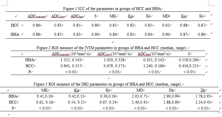
Figure.1 ICC of the parameters in groups of HCC and HHA
Figure.2 ROI measure of the IVIM parameters in groups of HHA and HCC (median, range).
Figure.3 ROI measure of the DKI parameters in groups of HHA and HCC (median, range).