2036
BOLD MRI of Activated Human PancreasBozhu Chen1, Jian He, and Zhengyang Zhou
1Radiology, Drum Tower Hospital, the Affiliated Hospital of Nanjing University Medical School, Nanjing, People's Republic of China
Synopsis
Glucose ingestion activates the pancreatic functions and increases the oxygen consumption, we hypothesized that BOLD MRI could detect the alterations during the glucose challenge. BOLD MRI was performed in 12 volunteers before and after glucose ingestion. A transient but significant decrease in pancreatic T2* values was observed after glucose ingestion. BOLD MRI may serve as a non-invasive tool to detect the activation of human pancreas.
Purpose
To test whether blood oxygen level dependent (BOLD) magnetic resonance imaging (MRI) can detect the activation of human pancreas.Methods
Healthy volunteers (n=12) were recruited for this pilot study and were asked to fast for 10 hours before examination. All volunteers were scanned in the supine position with 3.0 Tesla MR scanner (Ingenia; Philips Medical Systems, Best, The Netherlands) and a 32-element dS Torso phased-array surface coil. An axial T2*-weighted multiple gradient recalled echo (mGRE) sequence (flip angle, 27°; repetition time, 68 ms; field of view, 400 mm × 400 mm; matrix size, 256 × 256; section thickness, 5 mm; intersection gap, 1 mm) with ten echoes (4, 8, 12, 16, 20, 24, 28, 32, 36, and 40 ms) was performed for BOLD imaging. Oral intake of 50 mL 50% glucose solution was used to activate the pancreas. Scanning was done for 3 minutes at baseline and 10 minutes after glucose ingestion. T2* maps were generated by software built in the scanner, and pancreatic T2* values were obtained by place ROIs at each time point. To test whether a stimulus caused significant changes in the T2* values at a particular time point, the mean percentage of changes at each time point was compared to zero (the theoretical mean if no change had occurred) by Student’s t-test.Results and Discussion
A transient but significant decrease (-6.88 ± 1.01%) in pancreatic T2* values was observed within 5 min after glucose ingestion (Fig.1). The initial drop may indicate higher rate of oxygen consumption relative to blood flow as metabolic machinery become activated, which is similar to the liver[1]. It has been shown that glucose-induced O2 consumption may cause hypoxia in pancreatic beta-cells[2]. BOLD MRI enabled noninvasive quantification of pancreatic T2* changes during glucose stimulation. Further investigations are currently underway in order to confirm the application of this noninvasive technique in the assessment of pancreatic oxygenation and to explore its potential in the evaluation of the pancreatic function.Acknowledgements
No acknowledgement found.References
[1] Fan et al. (2006) ISMRM abs. 14:3249.
[2] Bensellam et al. (2012) PLoS One. 7: e29807.
Figures
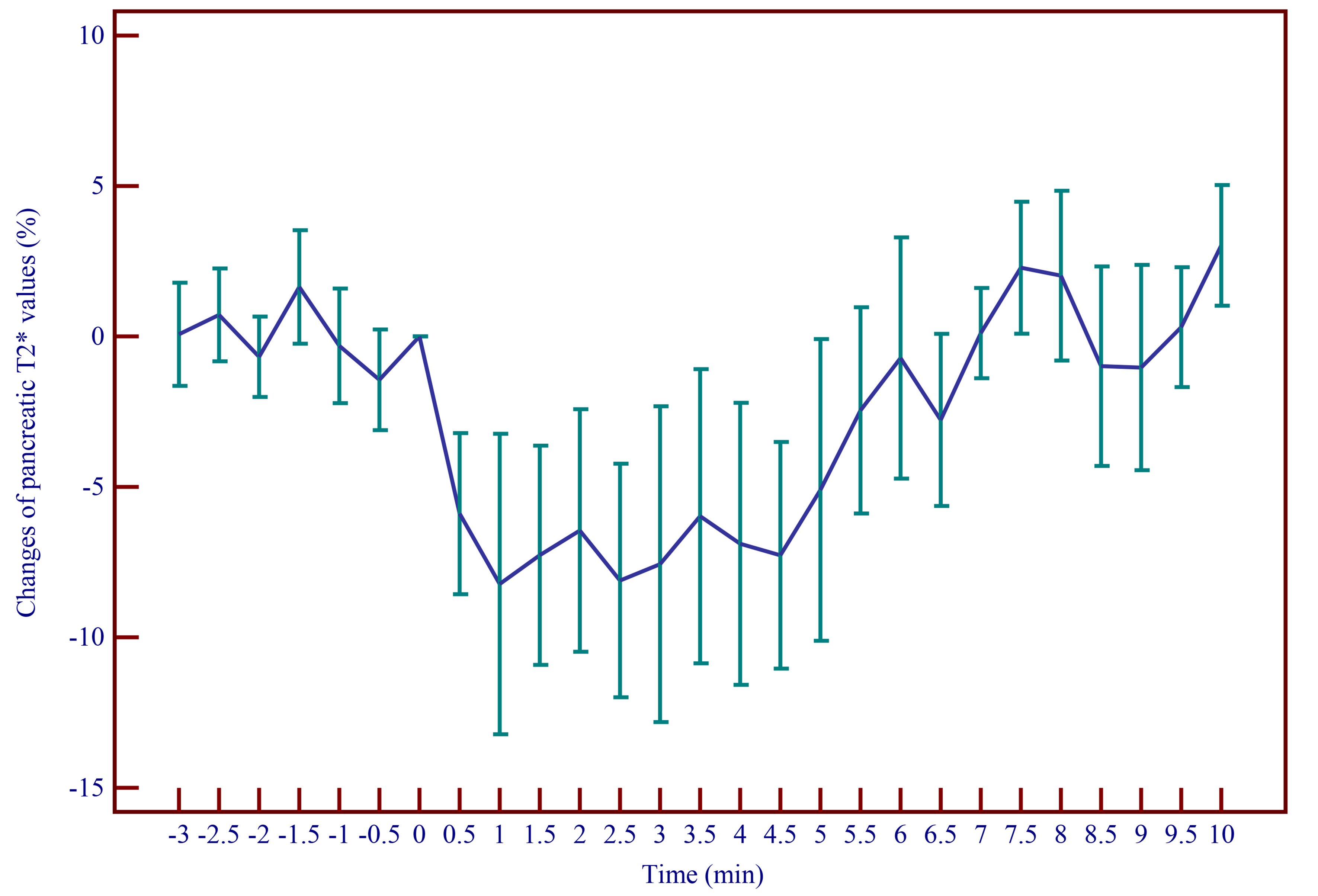
Figure 1. The graph shows normalized pancreatic T2* value
changes (mean of 12 subjects with 95% CI) before (-3 to -0.5 min) and after
(0.5 to 10 min) the ingestion of glucose.