2029
Liver T1rho detects liver fibrosis without impact of fatty liver in patients with chronic hepatitis B: a prospective study1Tianjin First Center Hospital, Tianjin, People's Republic of China, 2Philips healthcare, Beijing, People's Republic of China
Synopsis
This study investigated the merit of liver T1rho values for detecting fibrosis in patients with chronic hepatitis B and the potential impact of fatty liver on T1rho measurements. Eighteen healthy control subjects, eighteen patients with clinically diagnosed simple fatty and eighteen with liver fibrosis were underwent T1rho MRI and mDIXON-Quant. Mean T1rho values and fat fraction (FF) were compared among the three groups. Receiver operating characteristic (ROC) curve analysis was performed to evaluate the merit of T1rho values for detecting liver fibrosis. T1rho values were correlated with FF and clinical data. Our results showed significant differences in T1rho values among the three groups and T1rho had moderate diagnostic efficacy to detect fibrosis. T1rho values were not correlated with FF, subject age, or body mass index. We conclude liver T1rho values can be used as a MR biomarker for liver fibrosis and are not influenced by fatty liver severity.
Purpose
T1rho had a high sensitivity to low frequency motional processes, it can be used to investigate the macromolecular composition of and proton exchange within the tissues[1]. Liver fibrosis is the result of the biological deposition of macromolecules, so T1rho MR imaging may be sufficiently sensitive for evaluating liver fibrosis. Experimental studies in rats concluded that fibrosis degree was correlated with T1rho measurements and that T1rho MR imaging can be used to monitor liver injury and fibrosis degrees[2-3]. However, results from clinical studies were not consistent[4-5]. The purpose of this study is to investigate the merit of liver T1rho values for detecting fibrosis in patients with chronic hepatitis B and the potential impact of fatty liver on T1rho measurementsMaterials and methods
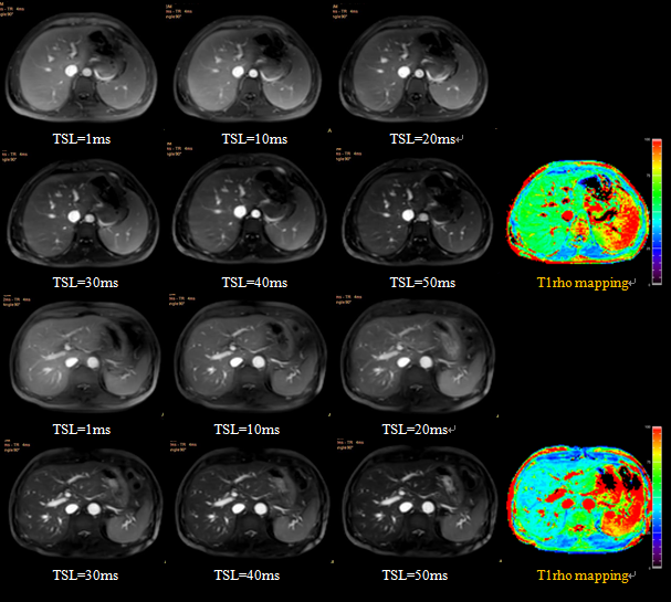
This prospective study was approved by the institutional ethics committee. Written informed consent was obtained. Healthy control subjects (n=18, F/M=5/13, mean age=51.8±10.3 years, range 34-74 years), patients with clinically diagnosed simple fatty liver (n=18, F/M=8/10, mean age=46.7±10.8 years, range 26-64 years), and patients with clinically diagnosed liver fibrosis (n=18, F/M=5/13, mean age=41.7±13.4 years, range 21-63 years) underwent T1rho magnetic resonance (MR) imaging and mDIXON-Quant collections. Liver T1rho and fat fraction (FF) values were measured by manually placing regions of interest. Mean T1rho values and FF were compared among the three groups. Receiver operating characteristic (ROC) curve analysis was performed to evaluate the merit of T1rho values for detecting liver fibrosis. T1rho values were correlated with FF and clinical data.Results
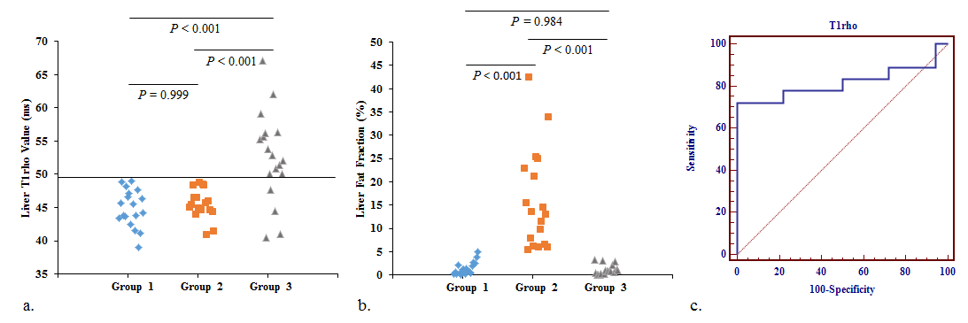
There were significant differences in T1rho values among the three groups (P < 0.001). Mean liver T1rho values in healthy control subjects (44.9 ± 2.8 ms) were similar to those of patients with simple fatty liver (45.0 ± 3.5 ms; P = 0.999) and significantly lower than those of patients with liver fibrosis (52.6 ± 6.8 ms; P < 0.001). Mean T1rho values performed well to detect fibrosis at a threshold of 49.5 ms (area under the ROC curve, 0.855). A significant difference in FF was also found among the three groups (P < 0.001). T1rho values were not correlated with FF, subject age, or body mass index (P > 0.05).Discussion
In this study, significant difference in liver T1rho values between normal control subjects and patients with liver fibrosis were seen. This finding highlights the potential value of T1rho for detecting fibrosis as indicated in previous studies. In addition, liver FF derived from mDIXON-Quant in both normal control subjects and patients with liver fibrosis differed from that assessed in patients with simple fatty liver, but no correlation between FF and T1rho values was found. This result suggests that the T1rho measurements were not influenced by fatty liver, enabling it to be used in more patients.Conclusion
Liver T1rho values can be used as a MR biomarker for liver fibrosis and are not influenced by fatty liver severity.Acknowledgements
No.References
1. Sepponen RE, Pohjonen JA, Sipponen JT, Tanttu JI. A method for T1ρ imaging. J Comput Assist Tomogr. 1985;9(6):1007-1011.
2. Zhao F, Wang YX, Yuan J, Deng M, Wong HL, Chu ES, et al. MR T1ρ as an imaging biomarker for monitoring liver injury progression and regression: an experimental study in rats with carbon tetrachloride intoxication. Eur Radiol. 2012;22(8):1709-1716.
3. Jiang JZ, Huang BS, Bin G, Chen SH, Feng F, Zou LQ. An experimental study on the assessment of rabbit hepatic fibrosis by using magnetic resonance T1ρ imaging. Magn Reson Imaging. 2016;34:308-311.
4. Singh A, Reddy D, Haris M, Cai K, Rajender Reddy K, Hariharan H, Reddy R. T1ρ MRI of healthy and fibrotic human livers at 1.5 T. J Transl Med. 2015;13:292.5. Takaymama Y, Nishie A, Asayama Y, Ushijima Y, Okamoto D, Fujita N, et al. T1ρ relaxation of the liver: a potential biomarker of liver function. J Magn Reson Imaging. 2015;42(1):188-195.
Figures