2027
Pre-operative MRI quantification of hepatic fat and its correlation with histology, BMI and length of peri-operative stay following resection of liver metastases1Department of Radiology, Memorial Sloan Kettering Cancer Center, New York, NY, United States
Synopsis
We correlate pre-operative quantification of hepatic fat on MRI in patients with colorectal cancer metastatic to the liver with the presence of fat on histology of the resected specimen, patient BMI and hospital length of stay.
Introduction
It is postulated that hepatic steatosis on preoperative MRI is an independent risk factor for complications following hepatic resection (1). Given the potential implication on post-surgical recovery and morbidity, we assess whether percentage fat quantification on in-phase and out-of-phase MRI correlates with the presence of steatosis on histological resection specimen, BMI, and the length of peri-operative hospital stay.
Methods
We reviewed a database of consecutive patients who underwent hepatic resection for colorectal liver metastases between April 2003 and March 2007. Inclusion criteria were patients with a baseline MRI as well as an additional pre-operative MRI. Patients were excluded from analysis if images were not digitalized. A total of 25 patients were analyzed, all of whom received pre-operative chemotherapy. For each study, a single slice in both the in-phase and the corresponding out-of-phase sequences was selected with 3 regions of interest measuring approximately 100 mm2 applied, taking care to avoid vessels, as well as a single ROI applied to the spleen. Fat fraction percentage was then calculated using the following formula (2):
$$〖Fat fraction〗_(CONVENTIONAL METHOD)= (〖T1_in_phase〗_LIVER/〖T1_in_phase〗_SPLEEN -〖T1_out_of_phase〗_LIVER/〖T1_out_of_phase〗_SPLEEN )/(2*〖T1_in_phase〗_LIVER/〖T1_in_phase〗_SPLEEN )$$
Results
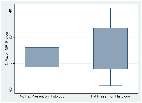
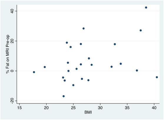
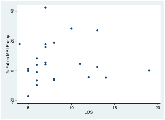
Baseline MRI fat fraction percentage shows a mean percentage fat in those with no fat on histology of 6.0% (SD 8.2) compared to those with fat on histology of 19.9 % (SD 20.1), p-value 0.01. Pre-operative MRI demonstrated a mean percentage fat in those with no fat on histology of 4.8 % (SD 10.2) compared to those with fat on histology of 10.6% (SD 24.0), p-value 0.01 (Fig. 1). Correlation coefficient for between BMI at time of surgery and the presence of fat on pre-operative MRI was 0.38 (Fig. 2). Correlation coefficient of length of stay with the percentage fat on pre-operative MRI was 0.02 (Fig. 3).
Discussion
As expected, pre-operative fat quantification on MRI best correlated with the presence of hepatic steatosis on histology. A weak correlation between BMI and percentage fat fraction was also found. Differences in correlation of percentage fat fraction with histology at baseline and pre-operative MRI possibly reflect chemotherapy effect. No correlation was found between length of stay and the percentage fat fraction calculated on pre-operative MRI, however patient sample size was small. Conclusion Pre-operative assessment of fat fraction percentage on MRI correlates with the presence of fat on histology, as well as with BMI. No correlation with peri-operative length of stay was found.
Acknowledgements
No acknowledgement found.References
1. d’Assignies G, Fayard C, Leitao H, et al. Liver steatosis assessed by preoperative MRI: An independent risk factor for severe complications after major hepatic resection. Surgery 2016; 159(4):1050-57.
2. Sirlin CB. Invited Commentary on Image-based quantification of hepatic fat: methods and clinical applications. Radiographics 2009; 29: 1277-80.
Figures