2007
Hepatocellular Carcinoma with Capsule Appearance in Gadoxetic Acid-enhanced MR Images : Correlation with Dynamic CT and Pathologic Fibrous Capsule1Radiology, Ajou University School of Medicine, Suwon, Korea, Republic of, 2Pathology, Ajou University School of Medicine, Suwon, Korea, Republic of
Synopsis
For visualization of capsule appearance on HCC, GaMR is comparable with dynamic CT. And pathologic fibrous capsule can be seen as hypointense rim in HBP. Lower visualization of capsule appearance in TP on GaMR seem to be parenchymal enhancement during the transitional phases. Hyperintensity rim in T2 weighted images with hopointense rim on HBP shows disruption of fibrous capsule with extension of tumor cells across the fibrous capsule in pathology. Hypointense rim on the HBP in the 2014 version of LI-RADS should not be considered as capsule appearance, and further study is also needed.
Purpose
The presence of a capsule appearance has often been considered a characteristic features of hepatocellular carcinoma(HCC) with favorable prognostic factor. The presence and recognition of a capsule appearance is important for the non-invasive diagnosis of HCC, and Liver Imaging Reporting and Data System (LI-RADS) included capsule appearance as a major feature. Recently, in the 2014 version of LI-RADS, various imaging features on hepatobiliary phase(HBP) were considered as ancillary features. The aim of this work was to establish the capability of gadoxeic acid-enhanced MR(GaMR) imaging to detect the presence of the capsule appearance and to correlate with dynamic CT and pathological findings.
Methods
Patients who underwent preoperative GaMR and dynamic CT between Jan 2013 and Dec 2015 were included for analysis. And 63 patients (54 males and 9 females, mean age 55.8years) were pathologically confirmed HCCs after surgical resection. Two observers independently reviewed presence or absence of capsule appearances on CT and GaMR images in the portal venous phase(PP) and delayed phase(DP)/transitional phase(TP). Hypointense rims surrounding nodules in HBP and T1- and T2-weighted images were considered as capsule in GaMR and correlated with patholgy. Interobserver agreement was assessed with kappa statistics, and consensus opinions were reached by conference. Pathological fibrous capsules were classified as complete, partial, and no fibrous capsule. Pathological fibrous capsules were compared and correlated with CT and GaMR, and sensitivity and specificity were calculated.Results
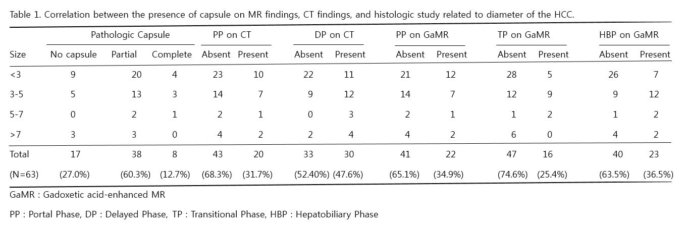
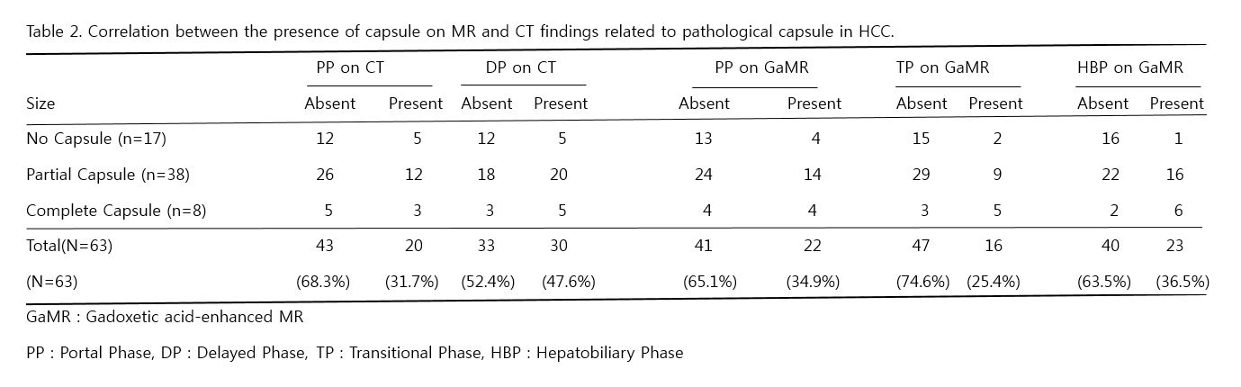
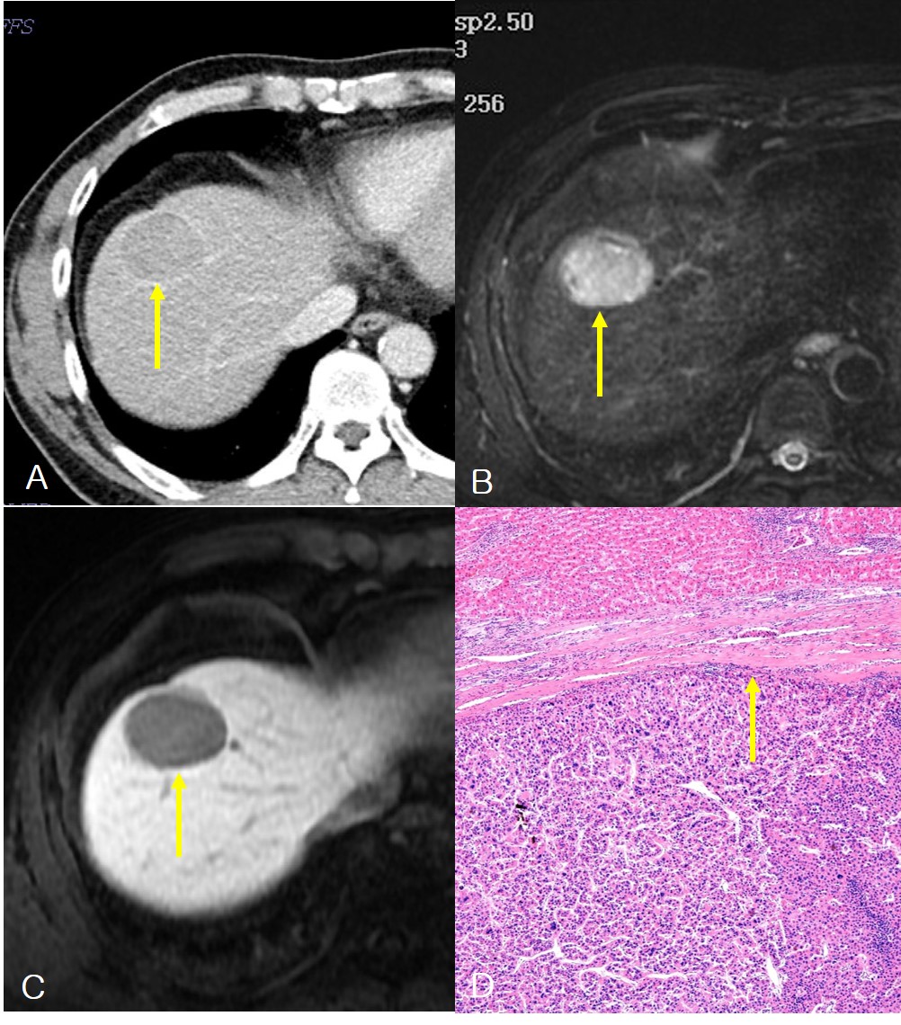
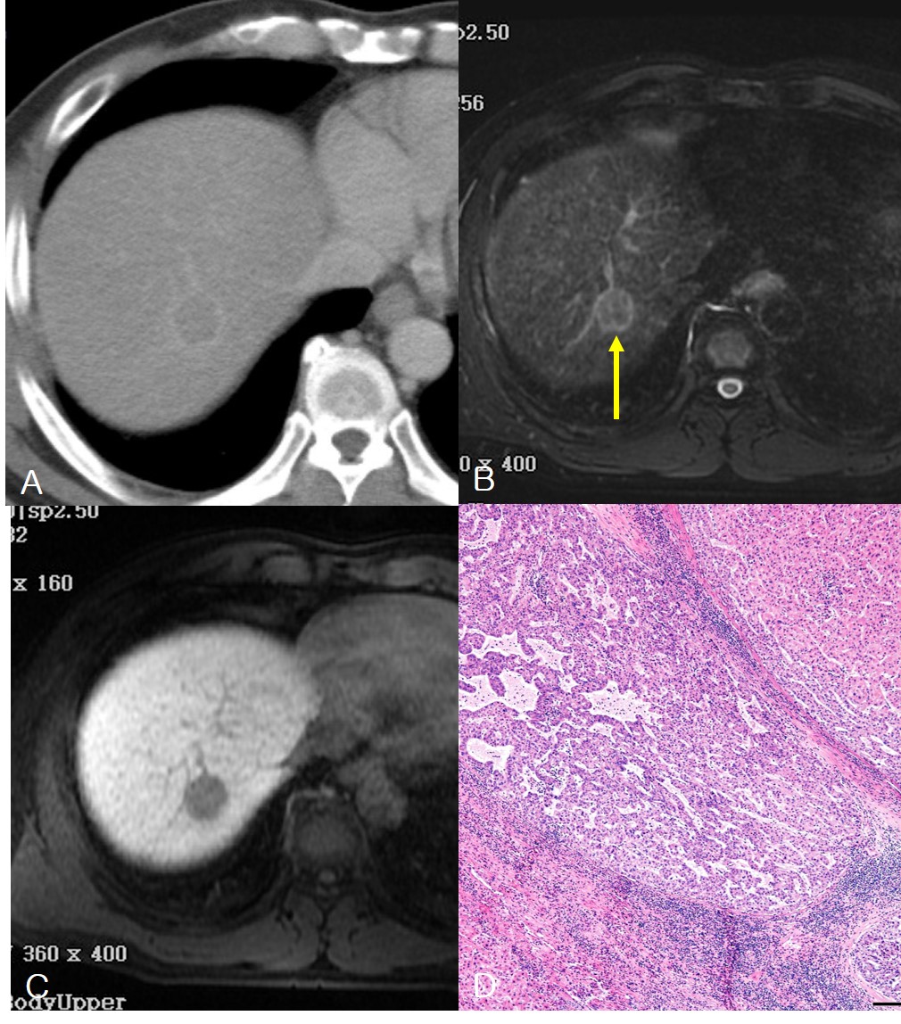
Interobserver agreement for the presence or absence of capsule appearance on GaMR was moderate on PP(k = 0.464; 95% confidence interval(CI): 0.265, 0.663) and HBP(k = 0.508; 95% CI: 0.303, 0.713), whereas fair in TP(k = 0.246; 95% CI: -0.0185, 0.511). Interobserver agreement for capsule appearance on CT was good on DP (k = 0.617; 95% CI: 0.423, 0.811) and moderate in PP(k = 0.555; 95% CI: 0.330, 0.781). Table 1 and 2 summarized presence or absence of capsule appearances on CT and GaMR with pathologic fibrous capsule. The capsule was histologically evident in 46 of 63 nodules (73.0%), complete in 8 (12.7%), and incomplete in 38(60.3%). Four cases(6.3%) were considered as pseudocapsule with false-positive fibrous capsule on CT and GaMR images. On GaMR, the capsule appearance was visible in 22(34.9%) in PP, 16(25.4%) in TP and 23(36.5%) on HBP(Figure 1). With CT, the capsule appearance was visible in 43(68.3%) in PP and 33(52.4%) on DP. Sensitivity for pathologic capsule was highest in DP on CT(54.4%) and specificity for pathologic capsule was highest in HBP on GaMR(94.1%). Four cases with hopointense rim on HBP shows high signal intensity rim around the HCC in T2 weighted images(Figure 2).Discussion
Hypointense rims surrounding nodules in HBP were considered as capsule in our study, and that correlated with fibrous capsule on pathology. Hypointense rim on the HBP in the 2014 version of LI-RADS should not be considered as capsule appearance, because other cause of hypointense rim on HBP should be considered and further study is also needed. Four cases with hopointense rim on HBP shows high signal intensity rim around the HCC in T2 weighted images. Pathologic correlation shows that disruption of fibrous capsule with extension of tumor cells across the fibrous capsule. Lower visualization of capsule appearance in TP on GaMR seem to be parenchymal enhancement during the transitional phases may mask the capsule appearance, as both the tumor capsule and enhancing adjacent hepatic parenchyma.Conclusion
For visualization of capsule appearance on HCC, GaMR is comparable with dynamic CT. And pathologic fibrous capsule can be seen as hypointense rim in HBP. Lower visualization of capsule appearance in TP on GaMR seem to be parenchymal enhancement during the transitional phases. Hypointense rim on the HBP in the 2014 version of LI-RADS should not be considered as capsule appearance, and further study is also needed.Acknowledgements
No acknowledgement found.References
1. Ishigami K, Yoshimitsu K, Nishihara Y, et al. Hepatocellular carcinoma with a pseudocapsule on gadolinium-enhanced MR images: correlation with histopathologic findings. Radiology. 2009;250(2):435-443.
2. Dioguardi Burgio M, Picone D, Cabibbo G, et al. MR-imaging features of hepatocellular carcinoma capsule appearance in cirrhotic liver: comparison of gadoxetic acid and gadobenate dimeglumine. Abdom Radiol. 2016;41(8):1546-1554.
Figures