1995
Hemodynamic Assessments of Hepatic Vasculatures using 4D-PCA and MRFD1Advanced Biomedical Imaging Research Center, Kobe University Graduate School of Medicine, Kobe, Japan, 2Center of Radiology and Radiation Oncology, Kobe University Hospital, Kobe, Japan, 3Radiology, Kobe University Graduate School of Medicine, Kobe, Japan
Synopsis
We introduced new assessment method of liver hemodynamics using 4D-PCA and new flow analytic technique including wall shear stresses. We found 4D-PCA and MRFD enables detailed hemodynamic assessment and has the potential to be used for liver disease assessments.
INTRODUCTION & PURPOSE
Liver has a unique hemodynamic system which is neither fully recognized nor understood.
Shear stresses to the liver and its vessels have been reported to play important roles to keep liver function and control its regeneration.
Non-contrast MRA using 4D phase-contrast angiography (4D-PCA) becomes clinically available and hemodynamic assessments using its cine data is reportedly useful for evaluations of cerebral and aortic aneurysms.
In this study, we applied these techniques to hemodynamic assessments of whole liver vessels.
The purpose of this study was to evaluate the capability of four-dimensional phase-contrast angiography (4D-PCA) and magnetic resonance fluid dynamics (MRFD) for assessment of hepatic vasculature.
MATERIALS & METHODS
Patients
25 patients (16 men, 9 women, mean 67.0 years), who were suspected to have hepato-biliary-pancreatic malignancy and underwent 3T-MRI, were enrolled.
The patients were divided into two groups.
•Liver disease group 9 HCCs, 5 liver metastases, 1 chronic hepatitis
•Non-liver disease group 4 pancreatic CAs, 1 papillary CA, 2 IPMNs、1 SPN, 1 GB stone, 1 cholangitis
MRI Technique
MR Units: Ingenia/Acheiva 3T (Philips Healthcare)
4D-PCA were obtained (TR/TE/FA: 4.1/2.4/10, matrix: 240x191 (ZIP256x256), FOV: 400mm, thk: 120mm, slice number: 60, voxel: 1.6x1.6x2.0mm, NEX: 1, PI: 3.0 (RL), 10 aqs/cardiac cycle, VENCs of 30 and 80 cm/s, scan time: 6-10min, pulse gating).
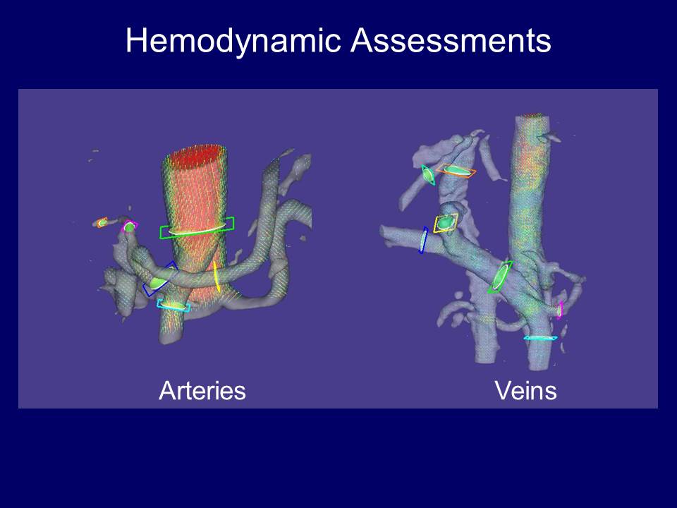
Hemodynamic Assessments
Hemodynamic assessment was performed on MRFD software (FLOVA, R'Tech).
•Abdominal Ao
•Celiac A, CHA, PHA, SPA, SMA
•Main, right, left portal veins, SMV, SPV
•Hepatic veins, IVC
Measured parameters
•Blood flow
•Blood velocity
•Wall Shear stress (WSS)
•Oscillatory shear gradient (OSI)
•Spatial WSS gradient (SWSSG)
•Gradient oscillatory number (GON)
Statistical Assessments
The hemodynamic values were compared
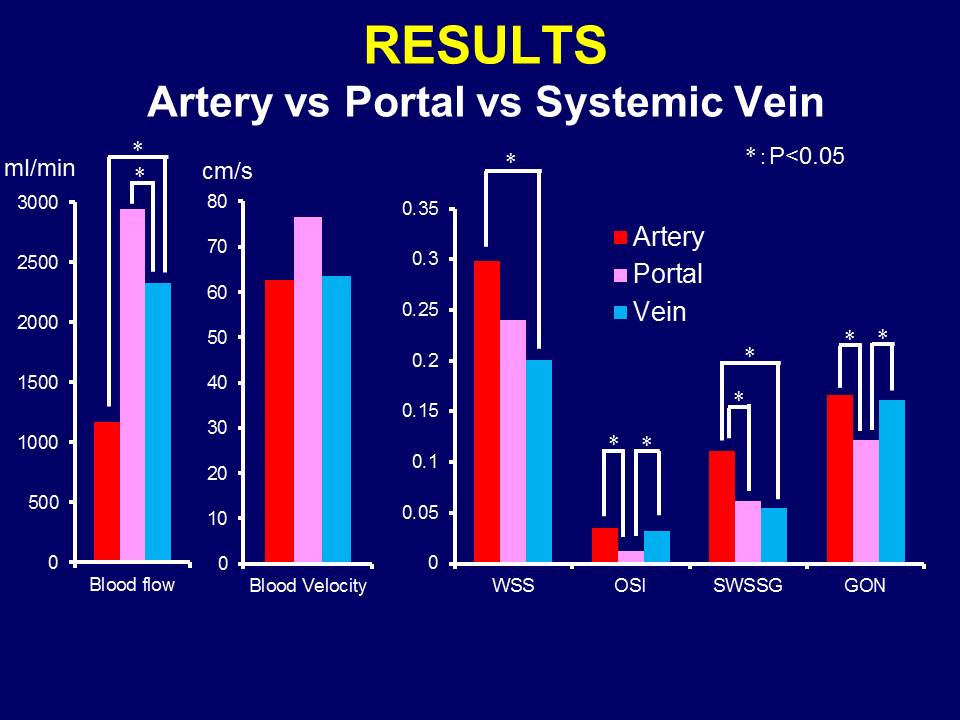
•among vessel types (overall artery, portal vein, and systemic vein)
•among arteries
•among portal veins
•among systemic veins
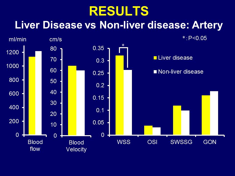
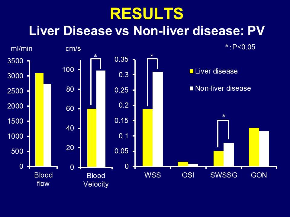
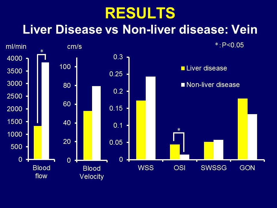
•between liver disease (n=15) and non-liver disease groups (n=10)
RESULTS
Hemodynamic assessment could be done in all the vessels except for proper hepatic arteries with diameters < 4mm.
Analysis time for each patient was roughly one hour.
Significant differences were found in flow and all 4 shear stresses in comparisons of vessel types (P<0.007), all parameters in comparisons of arteries (<0.004), flow and GON in comparisons of portal veins (<0.011), flow, WSS, and SWSSG in comparisons of systemic veins (<0.042), and systemic vein flow, portal velocity, arterial and portal WSSs, portal SWSSG, systemic vein OSI and GON in comparisons between the groups (<0.042).
DISCCUSSION & CONCLUSION
4D-PCA can be clinically used as a non-contrast angiography.
Further improvement of visualization is required for tiny abdominal vessels.
Hemodynamic assessments for each hepatic vessel could be done in relatively short time.
4D-PCA and MRFD enables detailed hemodynamic assessment and has the potential to be used for liver disease assessments.
Optimal assessment scheme is still unknown.
Acknowledgements
No acknowledgement found.References
•Mano Y, et al. Eur J Vasc Endovasc Surg 2013.
•Cheng CP, et al. Am J Physiol Heart Circ Physio. 2003.
•Stalder AF, et al. MRM 2008.
•Piva A, et al. Scand J Gastroenterol 2012.
Figures