1966
A Simple Phase Imaging REconstruction method (ASPIRE)1High Field Magnetic Resonance Centre, Department of Biomedical Imaging and Image-Guided Therapy, Medical University of Vienna, Vienna, Austria
Synopsis
Combining phase data from multi-channel coils at high field is challenging. Many approaches involve optimization or iterative steps which require offline reconstruction. We present a simple method that avoids the need for unwrapping or other fragile or time consuming steps by using echo times that satisfy $$$T_{E,2}=2\,\cdot\,T_{E,1}$$$. ASPIRE is compared with the Roemer method, MCPC-3D-I and the Hermitian inner product. ASPIRE achieves similar phase matching quality to Roemer but has the advantage that the combined phase values correspond to the B0 field. It is less computationally demanding than MCPC-3D-I and has higher CNR than the Hermitian inner product.
Purpose
The combination of phase images is a general problem at higher field strengths. The purpose is to develop a simple method for phase combination of multi-echo sequences which is suitable for implementation on a scanner.Theory
The general combination approach can be divided into three steps (Fig.1), which are common to a range of combination methods including the Roemer approach1 or MCPC-3D2:
1. Calculating the phase offset $$$\varphi_0$$$ for each coil element
2. Subtracting it from the individual coil element data
3. Combining the coil element data
The methods considered differ in the solving of Step 1 (Calculation of PO in Fig.1). In the Roemer approach, a separate reference scan from a volume coil is required to estimate the phase offsets. MCPC-3D-I requires a sequence with at least two echoes and performs unwrapping of all the single channel phases for the two echoes as a first step. From the unwrapped phase images, the phase offsets for each coil c can be calculated using the formula:
$$\varphi_0^c=\frac{T_{E,2}\,\varphi_{1,u}^c-T_{E,1}\,\varphi_{2,u}^c}{T_{E,2}-T_{E,1}}$$
As a final step, smoothing is applied to the calculated phase offsets to remove high frequency artifacts and noise.
ASPIRE uses a modified version of this formula that enables combination without unwrapping, if the echo times are chosen such that $$$T_{E,2}=2\,\cdot\,T_{E,1}$$$.
$$\varphi_0^c=\varphi_1^c-\frac{T_{E,1}}{T_{E,2}-T_{E,1}}\left(\varphi_2^c-\varphi_1^c\right)=\varphi_1^c-1\,\cdot\,PD\left(\varphi_2,\varphi_1\right)$$
The phase difference (PD) is calculated as the Hermitian inner product instead of for each coil individually, which makes the calculation more robust.
In general, scaling the phase values by an integer can be performed without introducing phase ambiguities other than multiples of 2π. This generalizes the echo time requirements to perform ASPIRE to
$$m\,\cdot\,T_{E,2}=\left(m+1\right)\,\cdot\,T_{E,1},$$
with m being an integer. The scaling factor m has to be applied to the HiP before subtracting it from the phase at the first echo time.
Methods
Data was acquired from a healthy volunteer with a 7T MR whole body Siemens MAGNETOM scanner with a 32-channel Nova Medical head coil, consisting of a birdcage transceive coil and 32 receive elements.
The target scan for phase combination was a high resolution multi-echo gradient-echo scan of the brain, which was acquired with monopolar readout in axial 3D, GRAPPA factor of 2 with 576x468x160 matrix, with 0.36mm in-plane resolution and 0.9mm thick slices, and a receiver bandwidth of 377Hz/pixel and TR=22ms. The sequence was acquired with 2 echoes with the echo times of 6.3ms and 12.6ms. As reference scans for the Roemer method two additional low resolution gradient echo scans have been acquired with a matrix size of 128x108x32, echo time of 4.6ms, one was acquired with the transceive volume coil and one was acquired with the 32 receive elements.
The ASPIRE combination was implemented in the Siemens image reconstruction environment (ICE). For comparison, the combination methods MCPC-3D-I, Roemer and Hermitian inner product (HiP)3 were performed on the high resolution data set.
Results
The combined phase images from ASPIRE and Roemer were compared to the HiP (Fig.2). The ASPIRE phase image accords closely to the HiP, differing only in having lower noise, while the Roemer phase image differs strongly from the HiP, indicating that it contains significant non-B0-related phase.
The quality of phase matching between the channels4 is indicated in Fig.3 and Fig.4, using following formula for calculation, which is applied voxel-per-voxel:
$$Q=\frac{\sqrt{\left|\sum{M_i^2\,\cdot\,e^{i\theta_c}}\right|}}{\sqrt{\sum{M_i^2}}}$$
The phase combination quality of ASPIRE and Roemer are equally good. The quality of MCPC-3D-I is high in most regions of the brain, however, in lower slices or disconnected regions the quality factor is low.
Table 1 shows the substantially higher CNR of ASPIRE and Roemer phase combination compared to a simple HiP phase difference, especially when combining both echoes.
The ASPIRE implementation in ICE requires a reconstruction time of about 30s on the scanner for the high resolution dataset.
Discussion
The Roemer method requires a measurement from a reference coil, which introduces additional susceptibility to motion and adds the transmit phase to the combined phase images. The main disadvantage of MCPC-3D-I phase combination is its dependence on unwrapping. ASPIRE, however, needs no reference scan and avoids the unwrapping to achieve a combination quality similar to that of the Roemer method. It also reaches a significantly higher CNR than the simple HiP approach. The requirement for applying ASPIRE is a multi-echo sequence in which two echoes fulfill the echo-time relation.Conclusion
The ASPIRE method features similar CNR to the Roemer approach without the need for an additional reference scan (volume coil) and removes all sources of phase from the combined image other than those originating in the B0-field. As such, it is optimal for use in Quantitative Susceptibility Mapping.Acknowledgements
This study was funded by the Austrian Science Fund (FWF) project KLI264.References
1. Roemer PB, Edelstein WA, Hayes CE, Souza SP, Mueller OM. The NMR phased array. Magn Reson Med 1990;16(2):192-225.
2. Robinson S, Grabner G, Witoszynskyj S, Trattnig S. Combining phase images from multi-channel RF coils using 3D phase offset maps derived from a dual-echo scan. Magnetic Resonance in Medicine 2011;65:1638-1648.
3. Bernstein MA, Grgic M, Brosnan TJ, Pelc NJ. Reconstructions of phase contrast, phased array multicoil data. Magn Reson Med 1994;32(3):330-334.
4. Robinson SD, Bredies K, Khabipova D, Dymerska B, Marques JP, Schweser F. An illustrated comparison of processing methods for MR phase imaging and QSM combining array coil signals and phase unwrapping, NMR in Biomedicine Early View 2016; http://dx.doi.org/10.1002/nbm.3601
5. Wu B, Li W, Avram AV, et al. Fast and tissue-optimized mapping of magnetic susceptibility and T2* with multi-echo and multi-shot spirals. NeuroImage 2012;59(1):297-305. http://dx.doi.org/10.1016/j.neuroimage.2011.07.019
Figures
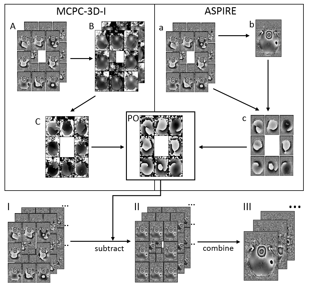
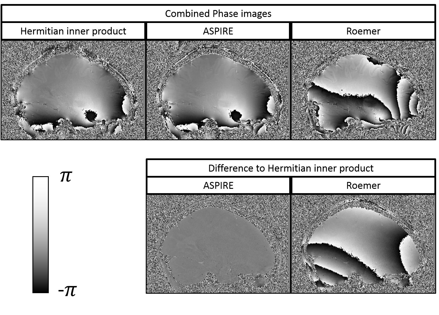
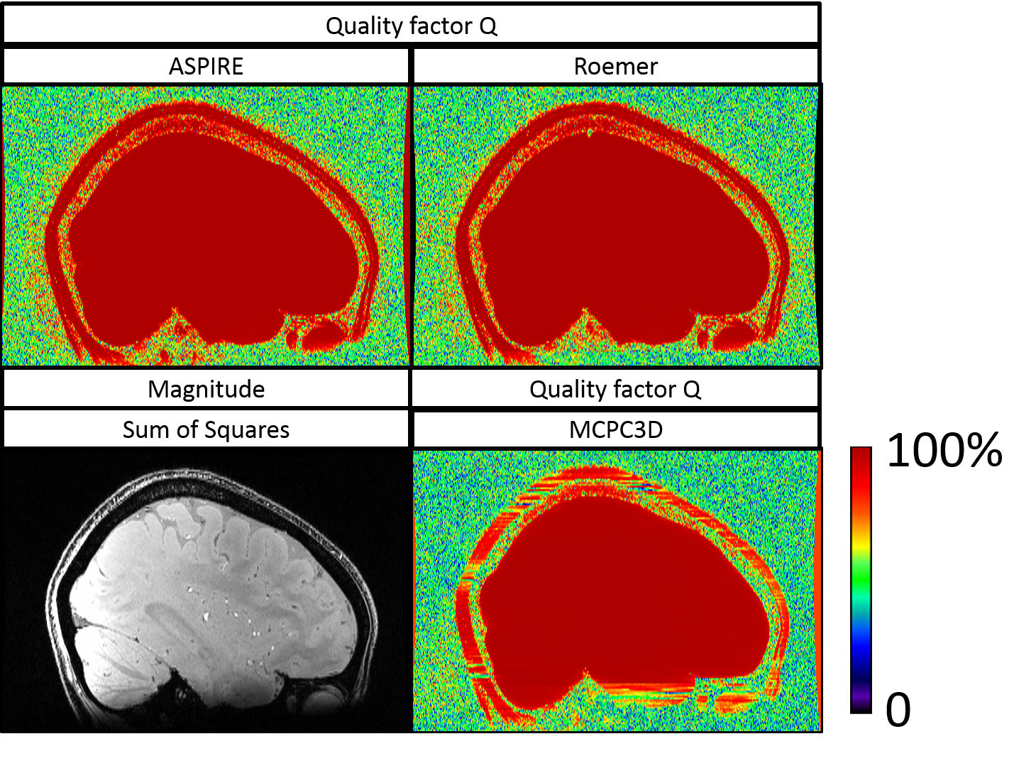

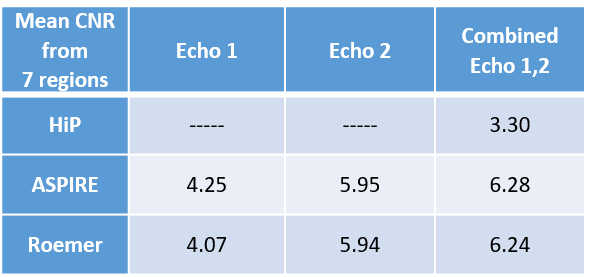
Table 1: CNR comparison for HiP, ASPIRE and Roemer; 5 sets of ROIs have been defined for measurement of the white-matter grey-matter contrast and two sets of ROIs have been defined for basal ganglia contrasts. The average CNR of the 7 regions is presented.
The HiP uses both echoes and therefore has only the combined average value. For the methods ASPIRE and Roemer, the CNR was calculated for each echo and for the combination, which was performed using weighting factors5. All CNR-values from ASPIRE and Roemer are substantially better than from HiP. ASPIRE and Roemer show very similar CNR values.