1868
Observing the evolution of MS plaques using SWI and DCS-PWI1Department of Radiology,First Affiliated Hospital of Dalian Medical University, Dalian 116011, People's Republic of China, 2GE healthcare China, Beijing
Synopsis
In this study,we aim to study the morphologic and micro-hemodynamic changes of MS plaques using SWI and DSC-PWI. We selected twenty-one MS patients diagnosed by the McDonald criteria (Revised Edition 2010) underwent MR scans including SWI and dynamical susceptibility contrasted MR perfusion weighted imaging (DSC-PWI) at baseline. Then Results were obtained by follow-up and scan. The MS plaques shows decreased phase value and blood perfusion. The characteristic of MS plaque is hypointense foci with small veins and only “abnormal vessels” region predict early changes.
Target audience
Any radiologists or neurologists interested in MS plaques evolutionPurpose
Based on the McDonald criteria (Revised Edition 2010)0[1],the temporal and spatial changes of the plaques in white matter on T2WI, T2 Flair or T1WI are the characteristic findings for MS patients. However, the occurrence of MS plaques before the hyperintense signal is observed on T2WI or T2 Flair. It has been proposed that the abnormal hemodynamic change may be one of cause of MS plaques formation [2].The phase of in this work, we aim to study the morphologic and micro-hemodynamic changes of MS plaques using SWI and DSC-PWI.Methods
Twenty-one MS patients (5 male, 16 female, mean of age=36.71 years old) diagnosed by the McDonald criteria (Revised Edition 2010) [1]underwent MR scans including SWI and dynamical susceptibility contrasted MR perfusion weighted imaging (DSC-PWI) at baseline. Follow up scans were then performed after interval from 2.0 months to 20.0 months (mean: 7.5 months) for all the subjects. All follow-up cases performed SWI and only 9 cases completed DSC-PWI scan. Features of MS plaques were assessed and classified in baseline and follow-up filtered phase image (FPI). Then numbers of MS plaques in FPI were also counted. The phase value and perfusion parameters (rCBF and rCBV) variation were also evaluated over time in the leisons.Results
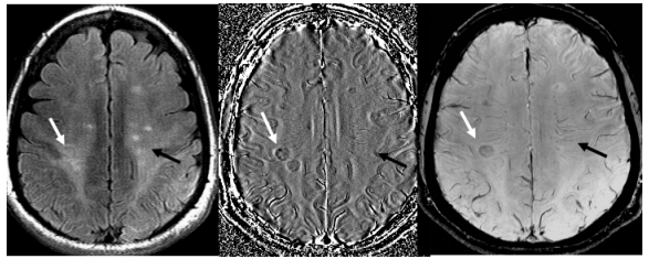
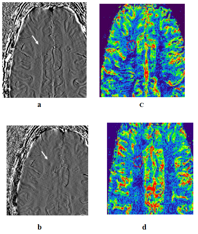
Images of a typical MS patient at baseline and at 7 month follow up are shown in Fig.1 and Fig.2 respectively. The measured phase, rCBF and rCBV values are summarized in Table1. In all cases 452 plaques were found in baseline T2 Flair and then only 211 plaques were seen in FPI. According to the relationship between the plaque and the small veins in the FPI, the MS plaques were divided (a total of 211) into 3 subtypes. TypeⅠ: The plaques in FPI shows the round or oval hypointense foci without small veins(77, 36%); TypeⅡ: The plaques in FPI showed hypointense foci with small veins(115, 55%); Type Ⅲ : The plaques in FPI showed only “abnormal vessels” region without hypointense foci(19, 9%)(Fig.1). Compared to those in NAWM, the phase values in plaques decrease, rCBF and rCBV also markedly decreased in baseline scan(P<0.01). In follow-up SWI cases, the numbers(211,205) and mean phase values of plaques were not significant different in FPI(P>0.05). In 9 follow-up DSC-PWI cases, the mean rCBF and rCBV did not change significantly over time (P>0.05). However, nine lesions evolved from only “abnormal vessels” region to hypointense foci with decreased phase value and rCBF and rCBV in 6 follow-up cases (28.57%, mean interval=7.0 months). Following up FPI (average 7 months), it was found that the “abnormal vessels” region decreased and disappeared, then there was a clear hypointense foci, which is from subtype III transitioned to subtype II, and even subtype I. Phase values in 9 plaques was significant related with rCBF and rCBV(r=0.79,P=0.01;r=0.75,P=0.02). (Table1,Fig.2)Discussion
In this study, only a small portion of the patients (6/21, 28.57%) with demyelinating lesions (9/452,2.0%) exhibiting "abnormal vascular" gradually evolved into hypointense foci. The blood flow perfusion (rCBF and rCBV) of these lesions also decreased to various extents. MR perfusion imaging studies have also confirmed that the brain tissue of MS patients is in a low perfusion state [3,4].The images of SWIMinIP and FPI of the MS in this study further confirmed the phenomenon that plaque and vascular inflammation are closely related. There was first discovered that only the “abnormal vessels” region, but no obvious low signal (19 , 9%) in the lesion area, which was the early change of the plaque formation. On the basis of the assumption of MS derived from chronic cerebrospinal venous insufficiency (CCSVI) by Zamboni [5], the author believes that the plaque in FPI was only "abnormal vessels" may be due to venous inflammation early, relative vascular expansion, the relative rise of blood flow and OEF corresponding increased.Conclusion
The MS plaques shows decreased phase value and blood perfusion. The variations from “abnormal vessels” region to hypointense foci in MS plaques may be the result of the disease progress. The characteristic of MS plaque is hypointense foci with small veins and only “abnormal vessels” region predict early changes. With the decrease of the blood flow perfusion, the deposition of iron in the plaque increased, and there was a trend towards the development of irreversible damage.Acknowledgements
No acknowledgement found.References
[1] Chris H. Polman, Stephen C. Reingold, Brenda Banwell, et al. Diagnostic Criteria for Multiple Sclerosis: 2010 Revisions to the McDonald Criteria:Ann Neurol,2011:69:292-302
[2]Ge Y, Zohrabian VM, Osa EO, et al. Diminished visibility of cerebral venous vasculature in multiple sclerosis by susceptibility-weighted imaging at 3.0 Tesla [J].J Magn Reson Imaging,2009,29(5):1190-1194
[3]Inglese M, Park SJ, Johnson G, et al. Deep gray matter perfusion in multiple sclerosis dynamic susceptibility contrast perfusion magnetic resonance imaging at 3 T. Arch Neurol,2007,64(2):196-202
[4]Law M, Saindane AM, Ge Y, et al. Microvascular abnormality in relapsing-remitting multiple sclerosis: perfusion MR imaging findings in normal-appearing white matter. Radiol,2004, 231(3):645–652
[5]Zamboni P, Menegatti E, Galeotti R, et al. The value of cerebral Doppler venous haemodynamics in the assessment of multiple sclerosis. J Neurol Sci,2009,282(1-2): 21–27
Figures