1866
Investigation of Diffusion Tensor Indices by TBSS analysis and of Patients with symptoms of neuropsychiatric systemic lupus erythematosus1Department of Radiology, Taichung Veterans General Hospital, Taichung city, Taiwan
Synopsis
In this project, we attempted to study the NPSLE subjects without abnormal lesion in conventional MR imaging in order to investigate the effective imaging biomarkers in early detection of neurological degeneration. Brain diffusion-tensor imaging with TBSS analysis was performed for studying the micro-structural alternations in NPSLE patients. The preliminary results illustrated statistically significant differences of FA and MD in some important nerve tracts between NPSLE patients and normal volunteers. There also existed a significant difference between NPSLE patients with and without depression.
Introduction
Systemic lupus erythematosus (SLE) is an autoimmune disease with multi-organ involvement. Once patient has neuropsychiatric signs and symptoms (eg. Stroke, movement disorder and psychosis), called neuropsychiatric SLE (NPSLE), the mortality and morbidity rates would be significantly increased. A variety of brain findings in NPSLE was presented, ranging from normal to predominantly global atrophy and focal or multiple nonspecific white matter disease. The underlying pathologic fact could be ischemia changes, vasculitis/vasculopathy , neuronal loss and demyelination. However, when it occurs, the prognosis is dismal. However, there was little known about pathogenic mechanisms leading to NP symptoms in SLE, whether it is caused by ischemia or auto-antibody mediated neuronal loss. Recent studies suggested that the change of indices indiffusion tensor images (DTI) is common in NPSLE [1]. Recently, a high prevalence of thecomorbidityanddepression of NPSLE was reported in the literatures.However, little is known about how these two different types of disease interfere with each other. This study aims to investigate white matter changes inpatients of NPSLE and normal volunteers usingdiffusion tensor imaging (DTI) and dedicated psychological tests.Methods
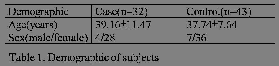
This study included 32 patients of NPSLE without obviously abnormal finding of morphological brain MRI and 43 normal volunteers. DTI data were acquired on a 1.5T Siemens MR system with following parameters: TR/TE=10000/107ms, b-value=1000 s/mm2, 30 directions, NEX=3 and voxel size=2*2*2mm3. Whole brain tracts of theFA and MD were carried out using FMRIB Software Library v5.0 (FSL)[2],andTract-Based Spatial Statistics (TBSS) [3]. TBSS pipeline was followed to show whole brain tract differences of both FA and MD in these two groups and P<0.05 for significance. Subsequently, partial Pearson correlation analyses were performed to correlate the clinicalevaluations with the regional DTI values within patient groups.Results
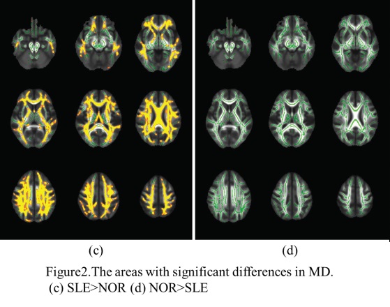
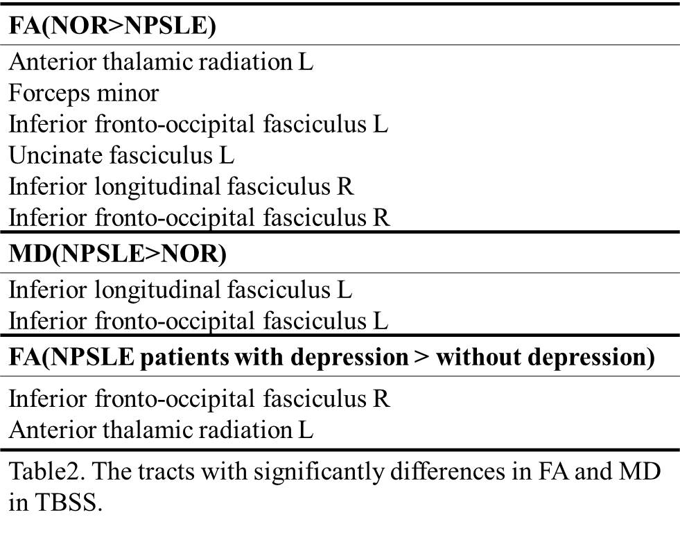
Demographic of case and control groups was showed in Table 1.Table 2 summarizes the results of DTI data from TBSS analysis that used permutation testing for group comparisons and FDR (5%) for correction of multiple comparisons with the threshold of p< 0.05 and at least 500 isotropic voxels. Left anterior thalamic radiation, forceps minor, bilateral fronto-occipital fasciculus, left uncinated fasciculus and right inferior longitudinal fasciculus had significantly lower FA in NPSLE patients than normal volunteers.There existed significantly mild correlation of impaired cognitive function in focus with FA in left fronto-occipital, right superior and inferior longitudinal fasciculus. A less number of nerve tracts, left inferior longitudinal and fronto-occipital fasciculus had significantly higher MD in NPSLE than the normal volunteers. There also existed significantly mild correlation of impaired cognitive function in language with MD in left inferior fronto-occipital and inferior longitudinal fasciculus. Figure 1 and 2are the t-statistics maps of FA and MD group comparisons overlaid on the MNI template in axial views. FA of left inferior fronto-occipital fasciculaus and left anterior thalamic radiation was significantly higher in NPSLE patients with depression than without depression.Conclusion
In this study, the results showed micro-structural alternations in NPSLE patients with grossly normal appearing brain, with lower FA and higher MD in several nerve tracts, which were mildly correlated with some specific cognitive function in focus and language . NPSLE patients with depression symptoms had usually been recognized as a worse clinical manifestation, but our results revealed symptom had higher FA in left inferior fronto-occipital fasciculaus and left anterior thalamic radiation. The finding would be much helpful for further investigation of the pathogenic mechanisms leading to NP symptoms in SLE.Acknowledgements
No acknowledgement found.References
1. Alexandra Popescu1and Amy H Kao. Neuropsychiatric Systemic Lupus Erythematosus. Curr Neuropharmacol. 2011 Sep; 9(3): 449–457.
2. Auning E et al.Neurobiological correlates of depressive symptoms in people with subjective and mild cognitive impairment.Acta Psychiatr Scand. 2015 Feb;131(2):139-47.
3. S.M. Smith, et al. Tract-based spatial statistics: Voxelwise analysis of multi-subject diffusion data. NeuroImage, 2006;31:1487-1505.
Figures
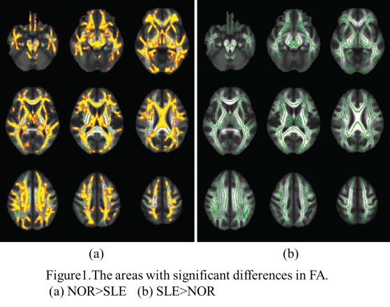
Figure1. The areas with significant differences in FA.
(a)NOR>NPSLE (b)NPSLE>NOR