1865
Functional Assessment of Lumbar Nerve Roots Using Direct Coronal Single-Shot Turbo Spin-Echo Diffusion Tensor Imaging - Application to Patients with Bilateral Spinal Canal Stenosis Showing Unilateral Neurological Symptom -1Radiology, Eastern Chiba Medical Center, Chiba, Japan, 2Philips Electronics Japan, Tokyo, Japan, 3Orthopaedic Surgery, Eastern Chiba Medical Center, Chiba, Japan, 4Kanazawa University, Ishikawa, Japan
Synopsis
Clinically, there are some patients with spinal canal stenosis who have unilateral neurological symptom despite the existence of bilateral nerve compression on the conventional MRI images. The purpose of this study was to investigate the availability of TSE-DTI for patients with bilateral spinal canal stenosis who have unilateral neurological symptom. At the level responsible for symptom, the average FA values of symptomatic side were significantly lower than those of asymptomatic side. FA values of TSE-DTI might be helpful in identification of responsible lumbar nerves roots for patients with bilateral spinal canal stenosis who have unilateral neurological symptom.
PURPOSE
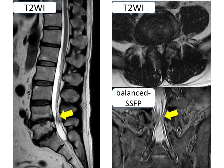
Diffusion Tensor Imaging (DTI) based on single-shot Echo Planner Imaging sequence (EPI-DTI) is established method to evaluate lumbar nerve roots compression, because several studies have shown that DTI and tractography of human lumbar nerves can visualize and quantitatively evaluate lumbar nerves by fractional anisotropy (FA)1,2. However, EPI-DTI has several problems such as long acquisition time and high geometric distortion. To solve these problems, we attempt to apply DTI based on single-shot Turbo Spin Echo sequence (TSE-DTI). Additionally, to reduce the total acquisition time, we applied TSE-DTI to set direct coronal acquisition. In previous study, we reported that TSE-DTI has a lower geometric distortion and it might more accurately evaluate compressed lumbar nerve roots compared to conventional EPI-DTI3 (Fig. 1). Clinically, there are some patients with spinal canal stenosis who have unilateral neurological symptom despite the existence of bilateral nerve compression on the conventional MRI images (Fig. 2). The purpose of this study was to investigate the availability of TSE-DTI for patients with bilateral spinal canal stenosis who have unilateral neurological symptom.METHODS
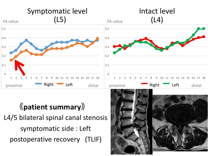
All subjects were examined with 1.5T whole-body clinical system (Ingenia, Philips Healthcare). The study was approved by the local IRB, and written informed consent was obtained from all subjects. Five patients with spinal canal stenosis, who have unilateral neurological symptom despite the existence of bilateral nerve compression on the conventional MRI images (median age, 69.8 years; range, 65–76 years), were examined by use of direct coronal TSE-DTI. FA values were continuously measured 18 points both proximally and distally to the bilateral lumbar foraminal zone at and adjacent to the levels responsible for symptoms (Fig. 3). We evaluated the relationship between average FA values and symptomatic side, and changes of the FA values in measurement point. Imaging parameters were; Coronal, voxel size=3.98× 3.98× 4.0mm3, FOV=350×350mm2, b-value=400s/mm2, MPG=32 directions, TR=3000ms, TE=49ms, and total acquisition time=6m25s.RESULTS AND DISCUSSION
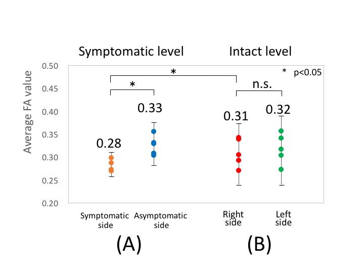
At the level responsible for symptom, the average FA values of symptomatic side were significantly lower than those of asymptomatic side (p<0.05) (Fig. 4). Also, the FA values of symptomatic side were lower than those of asymptomatic side on multipoint measurement (Fig. 5). Intraneural edema and demyelination caused by compression injury, was encouraged decreasing the FA values because of decreased diffusion anisotropy of the nerves4. Therefore, FA values of TSE-DTI might be helpful in the differential diagnosis for patients of bilateral spinal canal stenosis who have unilateral neurological symptom. The average FA values of asymptomatic level showed no significant differences in between right and left side. The FA values of distal points tended to higher than those of proximal points. Functional assessment of lumbar nerve roots with the FA values was required multipoint measurement, because FA values dynamically changed according to measurement points. CONCLUSION: FA values of TSE-DTI might be helpful in identification of responsible lumbar nerves roots for patients with bilateral spinal canal stenosis who have unilateral neurological symptom.CONCLUSION
FA values of TSE-DTI might be helpful in identification of responsible lumbar nerves roots for patients with bilateral spinal canal stenosis who have unilateral neurological symptom.Acknowledgements
No acknowledgement found.References
<1> BalbiV, et al. Tractography of lumbar nerve roots: initial results. Eur Radiol 2011;21(6):1153–9.
<2> Eguchi Y, et al. Quantitative evaluation and visualization of lumbar foraminal nerve root entrapment by using diffusion tensor imaging: preliminary results. Am J Neuroradiol 2011;32(10):1824–9.
<3> Sakai T, et al. Proc. ISMRM 2016:4477;
<4> Olmarker K, et al. Edema formation in spinal nerve roots induced by experimental, graded compression. An experimental study on the pig cauda equina with special reference to differences in effects between rapid and slow onset of compression. Spine. 1989 Jun;14(6):569-73.
Figures