1856
Mental training effects on adolescent brain networks1Radiology & Biomedical Imaging, University of California, San Francisco, San Francisco, CA, United States, 2Psychiatry, University of California, San Francisco, San Francisco, CA, United States, 3Clinical Neuroscience, Karolinska Institutet, Stockholm, Sweden, 4Clinical Sciences, Umeå Universitet, Umeå, Sweden, 5Psychology and Neurosciences Program, Stanford University, Stanford, CA, United States, 6PGSP-Stanford PsyD Consortium
Synopsis
In this study we used diffusion MRI network analyses to examine the effects of a 12-week training of attention and emotion regulation. Our preliminary results in 24 healthy adolescents demonstrate an improvement of executive attention and an increase of the node strength of the left anterior cingulate cortex.
Introduction
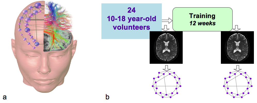
Executive attention and emotion regulation skills are important for a wide range of aspects in children's lives, and effective interventions aimed at their improvement need to be developed and assessed. The Training for Awareness, Resilience and Action (TARA) program initially developed for depressed adolescents uses a framework drawn from neuroscience, mindfulness, and yoga to promote attention and emotion regulation.1 The goal of this study was to assess the effects of the TARA training in healthy youth using MRI connectomics approach2 (Fig. 1a). Our hypotheses included: H1 - improvement of the executive attention, H2 - increase of the anterior cingulate cortex (ACC) node strength, H3 - decrease of depressive symptoms, H4 - increase of the caudate node strength.Methods
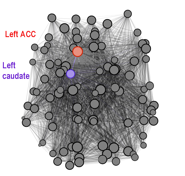
Participants were 24 healthy youth aged 10-18 years (mean age 15.43±2.13, 13F, 11M) recruited from the San Francisco Bay Area (Fig. 1b). The 12-week TARA training was delivered in groups of 6-10 participants. Pre-/post-training MRI scans were performed at a 3T GE MRI scanner and included a standard T1-weighted sequence and a Diffusion Tensor Imaging (DTI) sequence with TR=7.5s, minimum TE, FOV=25.6cm, 128×128 matrix, 2mm slice, resulting in a resolution of 2x2x2mm, 30 directions at b=1000s/mm2, ASSET acceleration factor=2, resulting in a scan time of 4min. The post-processing was performed using FSL, Diffusion Toolkit and Matlab and included eddy-current correction, DTI reconstruction and deterministic whole-brain streamline fiber tractography using FACT. T1-weighted data were registered to the b0-volume of the DTI dataset and to the MNI space template using FLIRT, allowing for application of the AAL atlas in the DTI space to produce 90 nodes of the network (Fig. 2). Weighted connectivity matrices were constructed using the AAL-based nodes and average fractional anisotropy (FA) within voxels passed by streamlines that connect every pair of nodes. The resulting networks were analyzed using the Brain Connectivity Toolbox.3 Pre-/post-training assessment of executive attention was performed in 23 participants using Attention Network Task (ANT).4 Pre-/post-training self-report measure of depressive symptoms (RADS-2)5 were collected in 17 participants.Results
The TARA training was
well received by the participants, as reflected by session-by-session
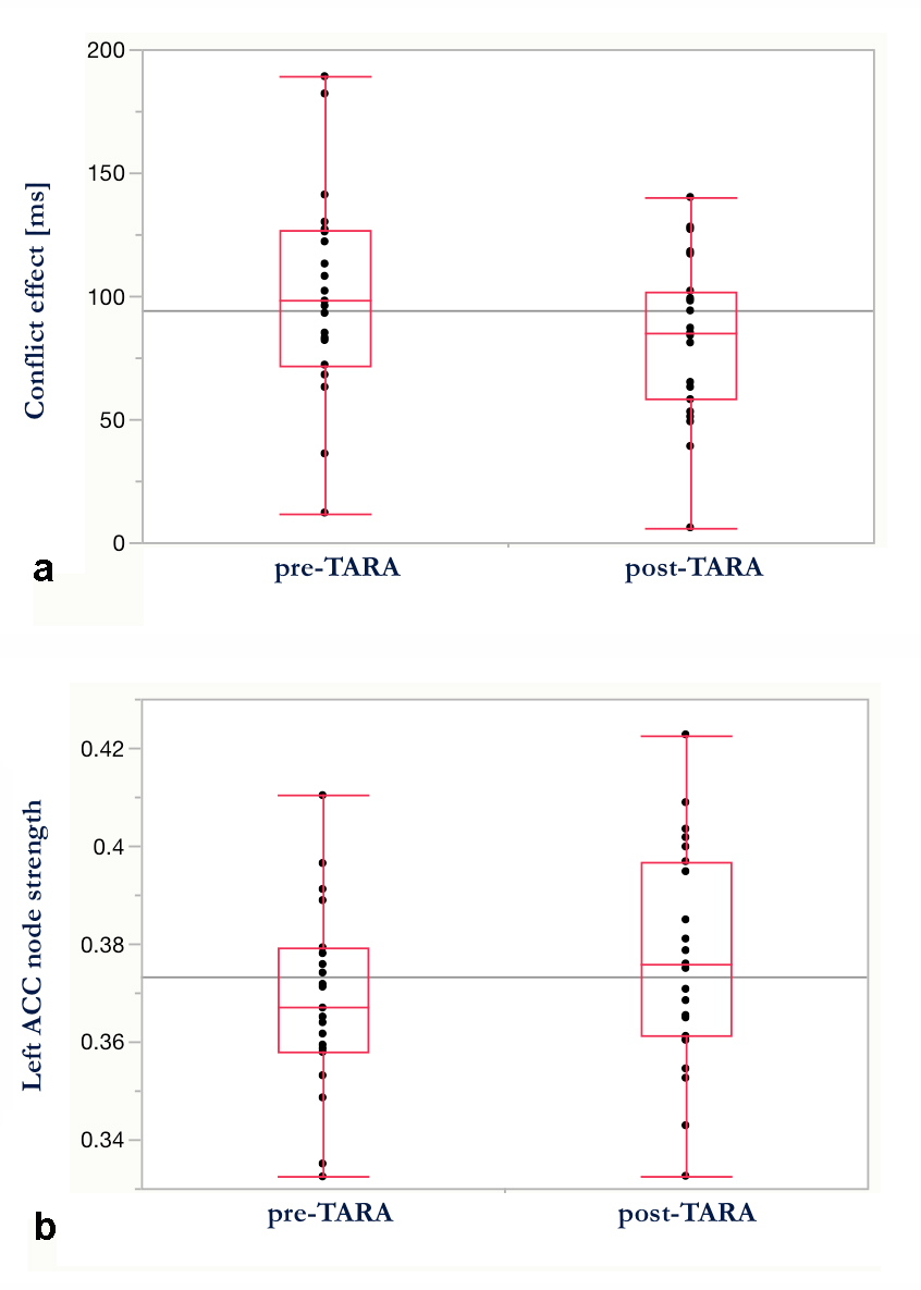
evaluations and feedback interviews. Executive attention of the participants
showed a significant improvement after the training (one-tailed paired t-test
assuming unequal variances, p=0.0236) (Fig. 3a). Network analysis revealed an increase
of the node strength of the left ACC (one-tailed paired t-test assuming unequal
variances, p=0.0326; excluding one outlier) (Fig. 3b). While depressive
symptoms did not show a significant change, there was an increase of the node
strength of the left caudate in participants with improved RADS-2 scores (N=7,
p=0.0336).Discussion
Our hypotheses H1 and H2 were confirmed, meaning an improvement of the executive attention and an increase of the ACC node strength, whereas depressive symptoms did not improve on average in this group of healthy youth. Those participants, whose depressive symptoms improved, did demonstrate an increase of the node strength of the left caudate. ACC is known to be involved in attentional processes; caudate is part of the reward circuit and has been implicated in adolescent depression.6 Potential cellular mechanisms of the FA-weighted network changes with training include: fiber reorganization, myelin formation, and myelin remodeling.7 To exclude possible maturational effects, future studies should include a control group. Further investigations are required to determine, which particular practices of the TARA training might be associated with the observed brain changes.Conclusions
Our results show that diffusion MRI-based brain network measures may be indicative of mental training effects in healthy adolescents.Acknowledgements
NIH R21AT009173, R01HD072074, UCSF Research Evaluation and Allocation Committee (REAC) and J. Jacobson Fund, and UCSF Radiology Seed #14-31References
1. Blom Henje E, Duncan LG, Ho TC, et al. The development of an RDoC based treatment program for adolescent depression “Training for Awareness, Resilience, and Action” (TARA). Front Hum Neurosci. 2014;8:630.
2. Hagmann P, Cammoun L, Gigandet X, et al. MR connectomics: Principles and challenges. J Neurosci Methods. 2010;194(1):34-45.
3. Rubinov M, Sporns O. Complex network measures of brain connectivity: uses and interpretations. Neuroimage. 2010;52(3):1059-69.
4. Fan J, McCandliss BD, Sommer T, et al. Testing the efficiency and independence of attentional networks. J Cogn Neurosci. 2002;14(3):340-347.
5. Reynolds WM. RADS-2, Reynolds Adolescent Depression Scale: Professional Manual. Psychological Assessment Resources, Lutz, FL. 2002.
6. Tymofiyeva O, Connolly CG, Ho TC, et al. DTI-based connectome analysis of adolescents with major depressive disorder reveals hypoconnectivity of the right caudate. J Affect Disord. 2016;207:18-25.
7. Zatorre RJ, Fields RD, Johansen-Berg H. Plasticity in gray and white: neuroimaging changes in brain structure during learning. Nature Neuroscience. 2012;15(4):528-536.
Figures