1854
Scan time reduction in DWI of the pancreas using simultaneous multislice technique with different acceleration factors: how fast can we go?1Department of of Diagnostic and Interventional Radiology, University Hospital of Tuebingen, Tuebingen, Germany, 2Section on Experimental Radiology, Department of Diagnostic and Interventional Radiology, University Hospital of Tuebingen, Tuebingen, Germany, 3Institute of Signal Processing and System Theory, University of Stuttgart, Stuttgart, Germany
Synopsis
We investigated the feasibility of simultaneous multislice-accelerated diffusion-weighted imaging of the pancreas using an acceleration factor of 2 and 3 (sms2/sms3-DWI) and its influence on image quality, acquisition time and apparent diffusion coefficients in comparison to conventional sequences (c-DWI) in ten healthy volunteers and 20 patients at 1.5 T. Images recorded with sms2-DWI offered high quality with a scan time reduction to one third; sms3-DWI showed significantly poorer overall image quality. In conclusion, sms2-DWI is feasible in clinical routine providing high image quality and a substantial reduction of acquisition time, whereas the use of higher acceleration factors is currently not recommended.
Purpose
To investigate the feasibility of simultaneous multislice-accelerated diffusion-weighted imaging (sms-DWI) of the pancreas with different acceleration factors and its influence on image quality, acquisition time and apparent diffusion coefficients (ADC) in comparison to conventional sequences.Methods
In this institutional review board approved study DWI of the pancreas was performed at 1.5 T in ten healthy volunteers (5 female, 5 male, mean age 29 years) and 20 patients (10 male, 10 female, mean age 59 years) undergoing clinically indicated MRI for the investigation of a suspected pancreatic pathology. In all participants sms-accelerated echo-planar diffusion-weighted imaging (provided by the University of Minnesota 1) with an acceleration factor (AF) of 2 (sms2-DWI) and 3 (sms3-DWI) and a conventional diffusion-weighted sequence (c-DWI) was performed. DWI was acquired in axial slice orientation in free-breathing. Diffusion gradients were applied in three orthogonal directions with b-values of 50, 400 and 800 s/mm². A monopolar diffusion gradient preparation scheme 2 and fat suppression with a spectral saturation pulse was used in all sequences. In all participants, the pancreas was screened for pancreatic lesions. For detailed image quality analysis following criteria were rated on a five-point Likert scale (5 = excellent): visibility of the pancreatic organ portions (head, body, tail), visibility of the splenic vein, presence of ghosting artifacts, interslice signal homogeneity and overall image quality. ADC values were recorded in a region-of-interest (ROI) analysis in head, body and tail. Qualitative and quantitative image parameters were compared using pairwise Dunn-Bonferroni post hoc test in case of significant difference in the overall Friedman test (p-values < 0.05 considered significant).Results
Sms-DWI of the pancreas was successfully performed in all volunteers and patients. Images recorded with sms2-DWI offered high image quality (overall image quality in b800: c-DWI: 4 [1; 5], sms2-DWI: 4 [1; 5]) with a scan time reduction to one third (c-DWI: 173 seconds, sms2-DWI: 56 seconds). Sms3-DWI showed significantly poorer overall image quality compared to c-DWI and sms2-DWI (sms3-DWI in b800: 3 [1; 5]; p < 0.0001). The existing pancreatic lesions showed comparable conspicuity in both c-DWI and sms2-DWI, while the generally poorer image quality in sms3-DWI resulted in a limited detectability of the lesions. ADC values in sms-DWI were generally lower compared to c-DWI with statistical significance comparing sms3-DWI to c-DWI in the pancreatic body and tail (body: c-DWI 1.4x10⁻³mm²/s, sms3-DWI 1.0x10⁻³mm²/s, p=0.028; tail: c-DWI 1.3x10⁻³mm²/s and sms3-DWI 1.0x10⁻³mm²/s, p=0.014).
Discussion
In the present study, the feasibility of simultaneous multislice-accelerated DWI of the pancreas was demonstrated in an experimental setting in volunteers as well as in patients scanned in clinical routine. Using an acceleration factor of 2, sms-DWI allowed considerable reduction of acquisition time while maintaining high diagnostic image quality and lesion conspicuity. When increasing the AF to 3, a significant deterioration in image quality was observed in both volunteers and patients with reduced visibility of all organ parts in the diffusion-weighted images and reduced image quality of the corresponding ADC maps. Moreover, artifacts and signal inhomogeneity in sms3-DWI were significantly increased, thus, discarding the use of higher AFs for clinical routine applications in its current implementation.
The ADC values of the pancreas measured in our study lie within the range of values reported in literature. 3,4 However, ADC values measured in sms-DWI were lower compared to c-DWI in all organ parts of the pancreas, an aspect having already been described using the sms-technique in DWI of the liver. 5 Possible reasons for this discrepancy include different noise statistics in the conventional and the sms sequences or variations in the signal processing of the coil channel combination between the two sequence techniques.
Yet, the clinical relevance of absolute ADC values is a matter of debate as a wide range of ADC values obtained by scanners provided by different vendors have been described. Thus, for now, relative values and visual interpretation seem more eligible for clinical use. 6
Acknowledgements
The authors would like to thank the University of Minnesota Center for Magnetic Resonance Research for providing continuous support regarding the sms MR sequence. Dr. Christian Wuerslin, Stanford, Radiological Sciences Laboratory, is gratefully acknowledged for kindly sharing with us the MATLAB post-processing software.References
1. Moeller S, Yacoub E, Olman CA, et al. Multiband multislice GE-EPI at 7 tesla, with 16-fold acceleration using partial parallel imaging with application to high spatial and temporal whole-brain fMRI. Magnetic Resonance in Medicine. 2010;63(5):1144-1153.
2. Stejskal EO, Tanner JE. Spin Diffusion Measurements: Spin Echoes in the Presence of a Time-Dependent Field Gradient. The Journal of Chemical Physics. 1965;42(1):288-292.
3. Herrmann J, Schoennagel BP, Roesch M, et al. Diffusion-weighted imaging of the healthy pancreas: ADC values are age and gender dependent. Journal of Magnetic Resonance Imaging. 2013;37(4):886-891.
4. Yoshikawa T, Kawamitsu H, Mitchell DG, et al. ADC Measurement of Abdominal Organs and Lesions Using Parallel Imaging Technique. American Journal of Roentgenology. 2006;187(6):1521-1530.
5. Obele CC, Glielmi C, Ream J, et al. Simultaneous Multislice Accelerated Free-Breathing Diffusion-Weighted Imaging of the Liver at 3T. Abdominal Imaging. 2015;40(7):2323-2330.
6. Sasaki M, Yamada K, Watanabe Y, et al. Variability in Absolute Apparent Diffusion Coefficient Values across Different Platforms May Be Substantial: A Multivendor, Multi-institutional Comparison Study. Radiology. 2008;249(2):624-630.
Figures
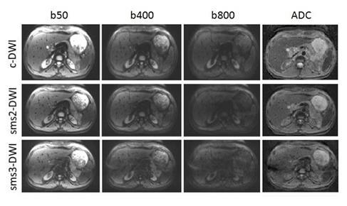
Figure 1 Image quality of conventional and simultaneous-multislice-accelerated DWI with different acceleration factors
Images obtained in a 31-year old female volunteer. Image set consisting of b50, b400 and b800 s/mm² and the corresponding ADC map. Diffusion-weighted images obtained with sms-accelerated DWI with an acceleration factor of 2 (sms2-DWI) and 3 (sms3-DWI) as well as the conventional sequence (c-DWI) as reference are shown. Windowing was optimized according to b-values. Image quality of sms2-DWI was rated equivalently high compared to c-DWI. In sms3-DWI, substantially reduced image quality was found with signal loss in the central image portions as well as pronounced ghosting.
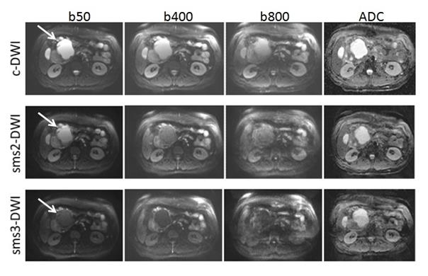
Figure 2 Image quality of conventional and simultaneous-multislice-accelerated DWI with different acceleration factors in a patient with a pancreatic pseudocyst
Images obtained in a 49-year old male patient with a history of chronic pancreatitis. Image set consisting of b50, b400, b800 s/mm² and the corresponding ADC map. Diffusion-weighted images obtained with sms-acceleration factor of 2 (sms2-DWI) and 3 (sms3-DWI) and the conventional sequence (c-DWI) as reference are shown. Windowing was optimized according to b-values. Image quality of sms2-DWI did not significantly differ from c-DWI, lesion conspicuity was equally high. In sms3-DWI, image quality and lesion conspicuity was substantially lower.
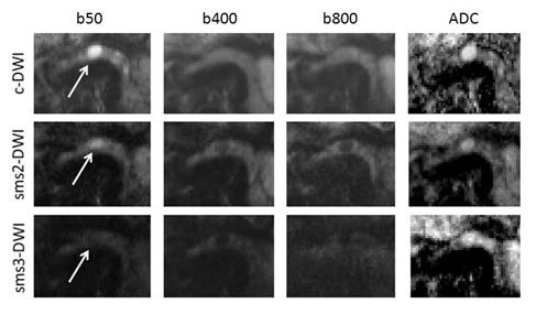
Figure 3 Image quality in conventional and simultaneous-multislice-accelerated DWI in a patient with a cystic lesion in the pancreatic body
Images obtained in a 74-year old male patient with a cystic lesion in the pancreatic body. Image set consisting of b50, b400, b800 s/mm² and the corresponding ADC map. Diffusion-weighted images obtained with sms-acceleration factor of 2 (sms2-DWI) and 3 (sms3-DWI) and the conventional sequence (c-DWI) as reference are shown. Image quality of sms2-DWI did not significantly differ from c-DWI. Lesion conspicuity was slightly reduced in sms2-DWI compared to c-DWI. In sms3-DWI, image quality and lesion conspicuity were substantially reduced.