1851
A longitudinal follow-up study of levator ani muscle injury during vaginal delivery using diffusion tensor imaging1Tianjin First Center Hospital, tianjin, People's Republic of China, 2Philips Healthcare, Beijing, China, beijing, People's Republic of China
Synopsis
Levator ani muscle (LAM) injury has been known to be highly associated with vaginal delivery, the natural recovery course of injured LAM is unclear recurrently. Diffusion tensor imaging (DTI) with fiber tracking is a useful noninvasive MRI technique, which can be used to assess pelvic floor muscles injury and recovery quantitatively, and the fiber tract can display the direction and thickness of LAM fibers. In this study, we tried to apply DTI imaging to explore the natural recovery course of LAM injury during vaginal delivery. And the results showed that the injured LAM during vaginal delivery has some degree of repair as time goes on.
Purpose
Levator ani muscle (LAM) injury has been known to be highly associated with vaginal delivery, the natural recovery course of injured LAM is unclear recurrently. Diffusion tensor imaging (DTI) with fiber tracking is a useful noninvasive MRI technique, which can be used to assess pelvic floor muscles injury and recovery quantitatively. The aim of this study is to to explore the natural recovery course of LAM injury during vaginal delivery using diffusion tensor imaging.Materials and methods
In this study, fifty primiparous women with six weeks after vaginal delivery(average age 27±2y) were included as the maternal group and seventeen age-matched nulliparous women volunteers(average age 26±1y) were included as control group . All the subjects underwent pelvic axial PDWI and DTI imaging with a 3T MRI scanner (Ingenia, Philips Healthcare, the Netherlands).The maternal group was divided into two subgroups: LAM injury group(fourteen cases) and non-LAM injury group(thirty-six cases) according to whether there was LAM injury evaluated with PDWI images. Then the LAM injury group was examined again after three months delivery as the follow-up group. Two radiologists measured FA and ADC values of LAM and observed the fibers direction and thickness of LAM on 3D fiber tracking images among LAM injury group, follow-up group and control group. Inter-rater reliability of each DTI parameters was evaluated using the intra-class correlation coefficient (ICC). FA, ADC values of LAM among the LAM injury group, follow-up group and control group were compared using one-way ANOVA and followed by post-hoc multiple comparison with Tukey method using IBM SPSS Statistics 21.0 (Armonk, New York, USA) and P<0.05 indicated a significant difference.Results
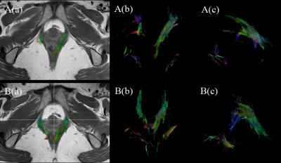
The inter-rater reliability between two radiologists for FA and ADC of LAM were good(ICC=0.75 and 0.92). For FA, the value of LAM in muscle injury group(0.27±0.04) was lower than the follow-up group(0.31±0.07) as well as lower than control group(0.33±0.07), there was a significant difference(P<0.005) among the three groups. Meanwhile, there was a significant difference between LAM injury group and control group(P=0.004). For ADC, there is no significant difference among LAM injury group, follow-up group and control group(1.40±0.24×10⁻³ mm2/s, 1.37±0.28×10⁻³ mm2/s, 1.33±0.24×10⁻³ mm2/s respectively, P=0.569). The LAM fibers in the LAM injury group are sparse and discontinuous. However, in the follow-up group, the LAM fibers are thickened and continuous, which were similar with those of control group.Discussion
A longitudinal follow-up evaluation of injured LAM recovery by using DTI was carried out in this study. The FA value of LAM in follow-up group was higher than that of LAM injury group, which indicates that the injured muscle diffusion direction is closer to harmonization during follow up and there were some degree recovery in the injured muscle. However, the difference of LAM ADC value among the three groups were not obvious, which may indicate the diffusion ability of injured muscle is unchanged as time goes on. Meanwhile, the fiber track images shows LAM fibers are more thickened and with a better direction in the follow-up group when compared with the LAM injury group(Fig.1).Conclusion
The injured LAM during vaginal delivery has some degree of repair as time goes on. DTI can be applied to assess the LAM injury and monitor its recovery course dynamically.Acknowledgements
No acknowledgement found.References
1.Can Cui, Yujiao Zhao, et al. Evaluation of the levator ani muscle in primiparas six weeks after vaginal delivery using diffusion tensor imaging and fiber tractography. International Society for Magnetic Resonance in Medicine, May 10, 2016. Singapore.
2.Rousset P, Delmas V, Buy JN, et al. In vivo visualization of the levator ani muscle subdivisions using MR fiber tractography with diffusion tensor imaging[J]. J Anat, 2012, 221(3): 221-228.
Figures
