1850
Distortion free whole-body diffusion weighted imaging using a self-adaptive post deformation algorithmLizhi Xie1, Bo Hou2, and Zhenyu Zhou1
1GE HealthCare, MR Research China, Beijing, People's Republic of China, 2Department of Radiology, Peking Union Medical College Hospital, People's Republic of China
Synopsis
Conventional Whole-Body DWI that consists of several sequential stations often troubled geometric distortions and signal drops in areas with strong susceptibility, such as the neck and lumbar vertebra. The aim of this study was to propose a new WBDWI protocol and post process deformation algorithm to get distortion free whole body images. Consistently good results were received and in helps to diagnose several cases of tumors that were previously unclear to the volunteers and patients.
Purpose
To develop an optimal protocol with a self-adaptive post deformation algorithm to access distortion-free Whole-body diffusion-weighted MR imaging (WBDWI).Introduction
Whole-Body DWI (WBDWI) allows the detection and characterization of preliminary lesions diagnosis and comprehensive tumor assessments in various malignant diseases [1, 2]. However, conventional WBDWI that consists of several sequential stations is frequently observed with geometric distortions and signal drops in those areas with strong susceptibility, such as the neck and lumbar vertebra. The aim of this study was to investigate the feasibility of an innovative WBDWI protocol and post deformation algorithm to achieve distortion-free WB images.Method
WBDWI acquisition requires several stations (4-8) due to the length of the magnet bore. In the conventional method, each station is regarded as a separate scan with a set of independent parameters including central frequency, signal gain and shimming values. The different parameter settings often result in image inhomogeneity and even distortions. On basis of our previous work [3], an innovative post algorithm was required to solve the observed serious deformation which were caused by metal influence and inhomogeneous magnetic field. The sagittal images were seriously deformed after the reconstruction of ax WBDWI. (Figure.1). Therefore, the post processing would focus on manipulating the sagittal position. Firstly, the positions of the spine (i.e. cervical, thoracic, and lumbar) were located. Based on the signal intensity curve, we can calculate the spine position of each station using the regional growth algorithm [4]. Secondly, the corrected position of the spine was achieved on each slice. The upper station was named as Sui and Sli for the lower station. The corrected position of spine in slice was calculated as j:$$$ C_{j}=S_u^i+(S_u^{i+1}+S_u^i-S_l^i-S_l^{i-1})*j/(2*SliceNumber)$$$. Lastly, - the images were resampled to achieve distortion-free sagittal images. The initial position of the spine was named as Si, and the corrected position of the spine as Sc, The value of after resampling was calculated using the formula: $$$V^{'}(x,y)=V(x,(y*S_{c})/S_{i})))(y<S_{C})$$$ (Figure.3).Experiment
All the healthy volunteers and patients underwent MR examination with Ready-fly WBDWI protocol, and the achieved images were analyzed and compared between two groups. The diffusion acquisition was achieved with a single shot EPI, and a 7-station setup was used to cover the whole body with the following parameters: axial plane, 28 slices with 6mm thickness a 0mm gap, FOV = 38cm×32cm, matrix = 160×192, TR/TE = 4600/52 ms, b value = 600 s/mm2, 4 averages. The scan time was 132 seconds for each station and overall 16 minutes for whole scanning session. To further test the robustness of this protocol, scanning with a low b value of 100 s/mm2 and a high b value of 800 s/mm2 were performed on the patient group.Results
Whole body DW images using conventional WBDWI and self-adaptive post deformation algorithm were achieved, where the post algorithm contributed to images with extremely eliminated distortions (Figure.2). No image distortion or inhomogeneity was observed and the metastases of the patient were clearly revealed with both b values. More experiments are required to investigate whether a low b value of 100 s/mm2 or a moderate to high b value of 800 s/mm2 will contribute to an overall image integrity that is better than the Ready-fly method.Discussion and conclusion
A new WBDWI post deformation algorithm was proposed, which m has been tested on 26 cases on 2 different sites to display consequently good results. This algorithm was able to assist with the diagnoses of several cases of tumors that were previously undefined for the subjects. Therefore, we proposed WBDWI to possess significant value in preliminary detection of metastatic tumors. Moreover, further tests will be performed to optimize the post algorithm on WBDWI.Acknowledgements
No acknowledgement found.References
[1]Horger et al, 2011, Acta Radiologica, 52:881-8.[2]Thomas C. Kwee et al, J. Magn. Reson. Imaging, 2014, Vol.40 (1).
[3]Xie Lizhi et al, 2015 ISMRM,1592
[4] Zhang Shuifa, et al. Transactions of the Chinese Society of Agricultural Engineering, 2013, volume 29(17):121-128(8).
Figures
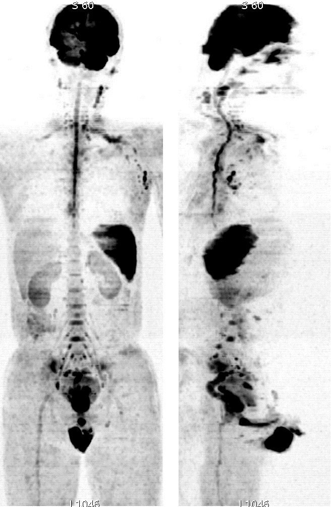
Figure1. Conventional WBDW of A
33yrs male volunteer
B =600 s/mm2 (Left:
coronal; Right: sagittal).
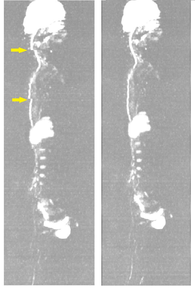
Figure2. Before and after self-adaptive post
deformation algorithm
A 38yrs male volunteer B =600 s/mm2 (Left: Ready-fly result;
Right: after post deformation algorithm).
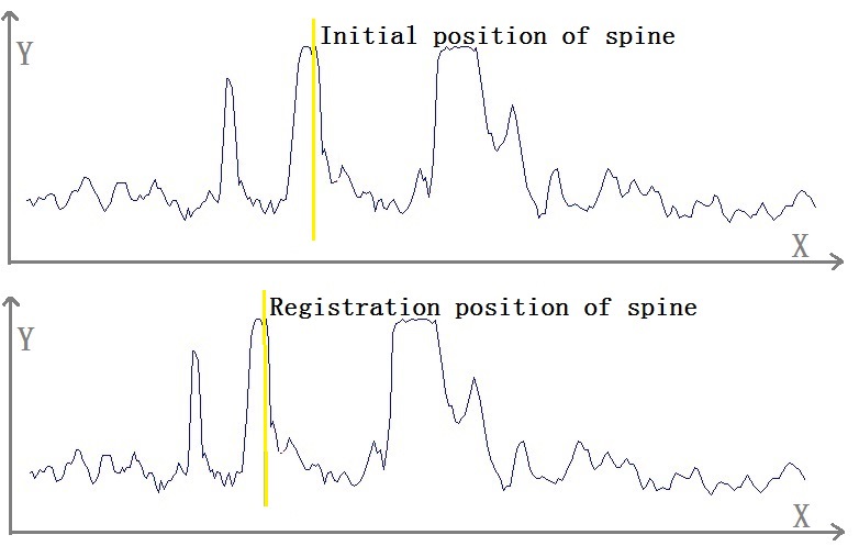
Figure3. Resampling algorithm.