1848
Myocardium Tissue DTI with Stimulated Echo at Large Susceptibility Induced B0 gradients: Examination of the Shimming Strategies Efficiency and Errors.Maxim Terekhov1, David Lohr1, and Laura Maria Schreiber1
1Comprehensive Heart Failure Center, University Hospital Wuerzburg, Wuerzburg, Germany
Synopsis
In this paper, we investigated experimentally and statistically the effect of distortions of myocardium DTI with STEAM-EPI due to susceptibility induced gradients varied in a range of factor 10 to 20 to the reference. The special focus was given to examining the effect of prolonged EPI-readout, B0-shimming effect and motion-induced shimming errors relevant for high-resolution DTI in-vivo. Fresh ex-vivo pig hearts were used for DTI measurements with an in-house developed STEAM-EPI sequence. The distribution of diffusion directions components was found well preserved for prolonged readout even at high internal gradient.
Introduction
The diffusion tensor imaging plays important role in cardiac MRI providing information on both structural and functional properties of the myocardium tissue1. To deal with the relatively short T2*-time of myocardium the stimulated echo-based sequences (STEAM) is typically used to provide sufficient time for the diffusion encoding by splitting it between two RR-intervals. To cover enough diffusion directions within single breath-hold an EPI-readout is normally employed. Both features make STEAM-EPI cardiac diffusion measurements extra sensitive to the B0-inhomogeniety, which are intrinsically strong within the heart due to heterogeneous structure and large blood volume. Additional susceptibility gradients are generated by the proximity of low density lung tissue. Therefore, the proper shimming strategy is crucial. The informativity of cMRI is typically limited by the low spatial resolution and SNR increasing the interest in using the ultra-high B0 for the high resolution cardiac DTI. In this paper, we investigated the effect of distortions of myocardium DTI with STEAM-EPI due to susceptibility induced gradients typical for high B0 fields. The special focus was given to examining the effect of B0-shimming effect and motion-induced errors relevant for the in-vivo.Materials and Methods
Fresh ex-vivo pig heart (n=3) were used for DTI measurements with an in-house developed STEAM-EPI sequence. To provide difference in the B0-homogeniety the containers with hearts were filled with 0.9% NaCl solution or left empty (referred further as “water” and “air” measurements, respectively). Measurements were done using 15-channel knee coil on 3T Siemens Magnetom “Prisma” scanner. The STEAM mixing time was limited to 250ms to provide sufficient SNR. The TE was varied from 45ms to 75ms (6/8 partial Fourier, iPAT=0) to simulate the potential for increasing spatial resolution. Matrix size=128x128, FOV=205mm, pixel bandwidth=1560Hz, 6 directions diffusion encoding were used. For the 3mm slice, the 15mm and 50mm shimming volume slabs were set. Shimming error due to heart motion were simulated by shifting the shim slabs as demonstrated by Figure 4(a). The whole heart 3D B0-maps with isotropic 1mm2x1.5 mm voxels were built using phase maps acquired by 3D GRE with TE=4.7 and 9.6ms. The standard scanner-side DTI reconstruction of diffusion tensor (ADC, fractional anisotropy, eigenvalues and main eigenvector (E1) components) were used. The analyzed parameters were: 1) relative bias (%) of isotropic diffusion coefficient (∆ADC) between TE=45ms and 75ms and its interquartile range (IQR) 2) similarity of the E1 components structure characterized by relative cross-correlation of its probability density functions (PDF), where 100% corresponds to identical PDFs.Results
Figure 1 shows the difference in B0-gradients maps in the fresh ex-vivo pig heart surrounded by physiological solution and air. The dramatic increase of the B0-heterogeneity in the air due to susceptibility gradients is observed. Figure 2 demonstrates the measured ∆ADC due to prolonged EPI-readout time (TE). The increase of the ∆ADC IQR by factor 1.5-2 is observed for the air in comparison with the water. Figure 3 shows that the increase of B0-heterogeneity in the air leads to essential distortions in characteristic segmental structure of main diffusion directions distribution within myocardium. Figure 4 demonstrates the combined effect of B0 heterogeneity and “off-slice” shimming error for the ADC and diffusion direction structure integrity.Discussion and Conclusion
The increase of the B0-heterogeneity by factor 10 to 20 in the “air” in comparison to “water” environment by susceptibility gradients may be considered as representative, for example, for the in-vivo measurements of the human and large animal heart at 7T B0 field. As expected, the additional shortening of T2* is manifested via heterogeneity of the bias of ADC for increased TE. This, in turn leads to distortion of both STEAM encoding and measuring the diffusion parameters by EPI. The distortions of E1 directions distribution in the air is well recognized on Figure 3 (top) both visually and in PDFs of individual components (bottom panels). However, the increase of TE up to 75ms does not change significantly the E1 direction structure even in the air. Moreover, the similarity of PDFs is very high in the air. Later finding is important for the high-resolution DTI where long TE time is mandatory. Up to 25% increased heterogeneity of the ADC-bias due to longer EPI readout (b) is observed. The relative similarity of the E1 components distribution drops down for the “off-slice” shimming but not for the shimming slab of 50mm. This means that “global” shimming not focused to the specific slice should be preferable for the high B0 gradients if standard scanner algorithms are used. Alternatively, the dynamic shimming with the navigation to the measured slice should be applied.Acknowledgements
We acknowledge financial support of German Ministry of Education and Research (BMBF), grants: 01EO1004, 01E1O1504.References
1. Choukri Mekkaouia, Timothy G. Reesea, Marcel P. Jackowskib,Himanshu Bhatc and David E. Sosnovik, Diffusion MRI in the heart, NMR Biomed: 2015; Epub doi: 10.1002/nbm.3426Figures
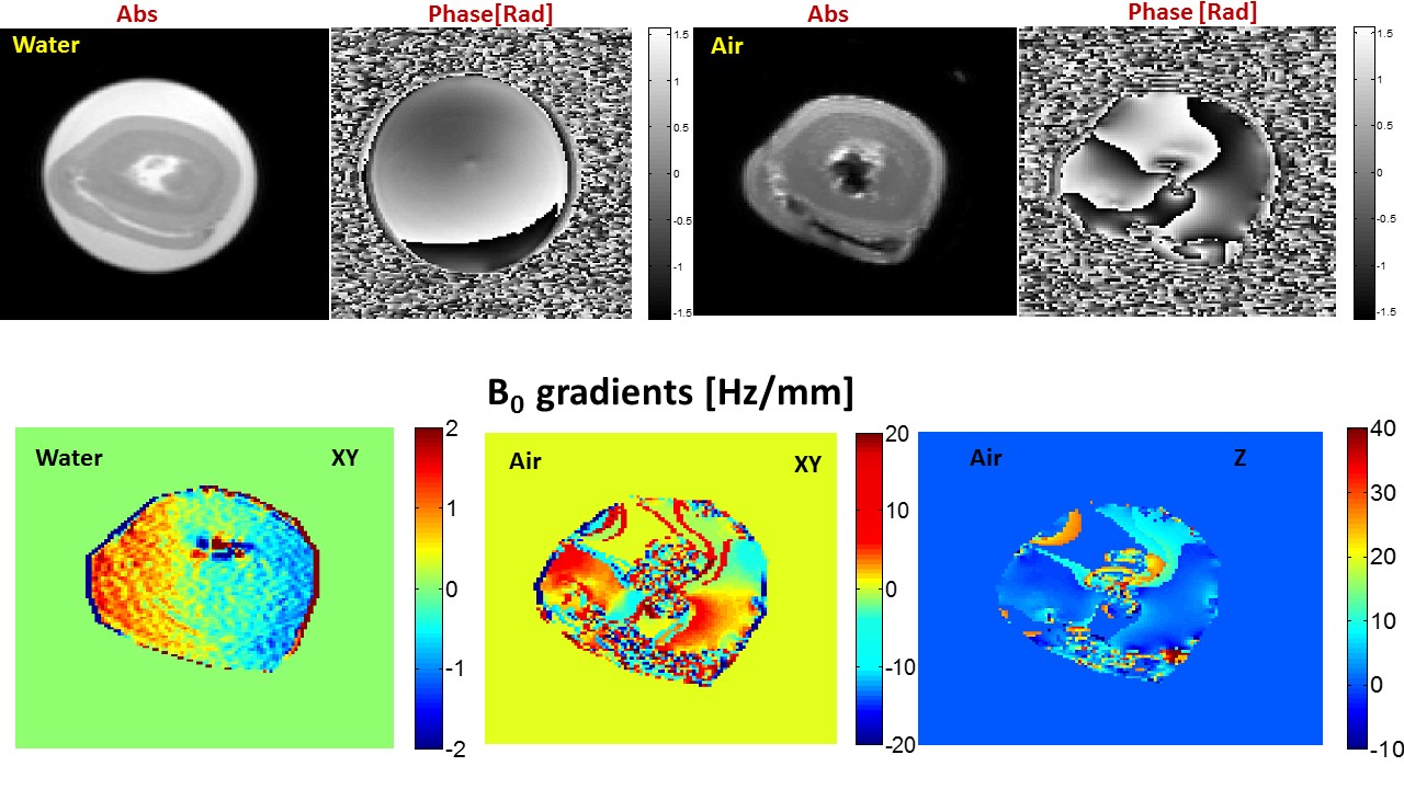
Figure 1 B0 gradients maps measured at 3T in the fresh
ex-vivo pig heart myocardium surrounded by physiological solution and air after
applying standard scanner “cardiac” 3D shimming. The dramatic increase (factor 10 to 20) of
the B0 heterogeneity in the “air” in comparison to “water” environment
due to susceptibility induced gradients may be considered as representative for
the in-vivo measurements of the human and large animal heart at 7T field.
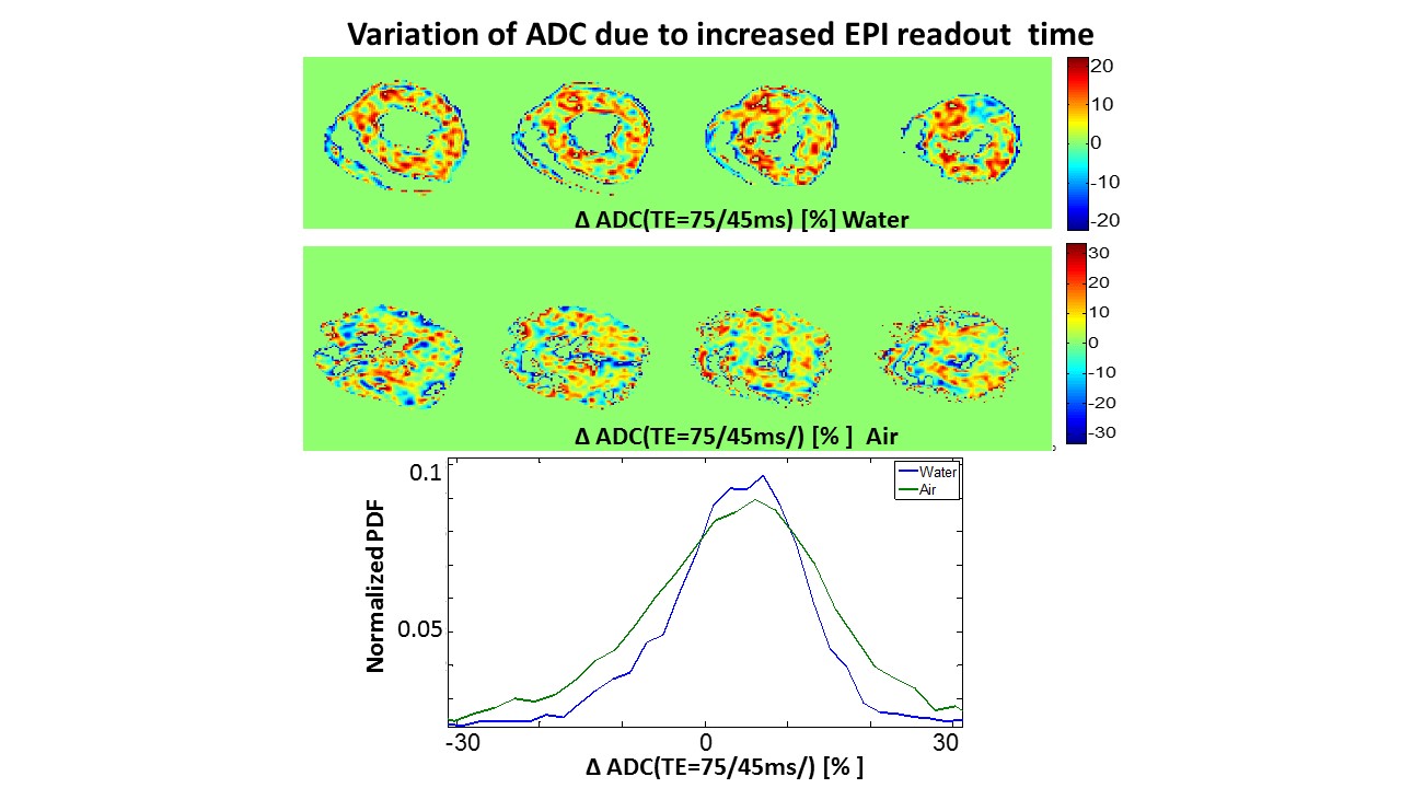
The
intrinsic B0 gradients leads to systematic bias of ADC by the
increased EPI readout time (TE). Thе bias becomes additionally more heterogeneous with
the B0 field. The increase of the ADC interquartile range by factor
1.5-2 was observed for measurements in the air (“poor shimming”) in comparison
with the one in water environment (“good shimming”).
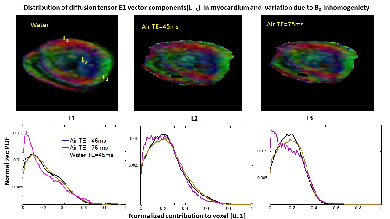
The heterogeneous B0
leads to problems of proper encoding and measuring of diffusion tensor
parameters. The visualized directional structure (shown by conventional DTI
color coded diffusion tensor main eignenvector components L1-3)
suffers from both ADC bias and geometry distortions by “air” measurements. One may notice, however, that additional
increase of EPI readout time from 45 to 75ms does not change significantly the
visualized structure and histograms shape is very well preserved (bottom
panels). Later finding is important for
the high-resolution DTI where long TE time is mandatory.
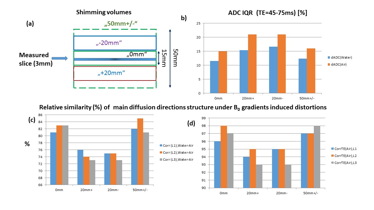
The effect of the “off-slice”
shimming for ADC and diffusion directions is expected due to heart motion and
breathing by small shim volume focused to measured slice. The scheme of the shimming
volumes positioning relative to measured slice is shown on (a). Moving shim volume out
of measured slice leads to increased heterogeneity of the ADC bias due to
longer EPI readout (b). The integrity
of the directions distribution(characterized by the relative cross-correlation of probability density functions for each direction component) decreases due to off-slice shimming ("+" and “-20mm”). The "50mm slab" restore the situation back to the “in-slice” shimming.