1843
Intravoxel Incoherent Motion (IVIM) in Evaluation of Orbital Masses1Department of Radiology, Renmin Hospital of Wuhan University, Wuhan, People's Republic of China, 2GE Healthcare China, Shanghai, People's Republic of China
Synopsis
Diffusion-weighted imaging (DWI) has been proven that malignant orbital masses demonstrate significantly and visually appreciable lower apparent diffusion coefficient (ADC) than benign orbital masses[1-2]. Nevertheless, ADC cannot separate the pure molecular diffusion from the motion of water molecules in the capillary network; thus, perfusion contamination would increase the ADC value. According to the IVIM DWI model[3], both microscopic perfusion and diffusivity can be separated using a biexponential decay function, providing additional parameters for tissue characterization. From the result we can see that it is feasible that quantitative parameters of orbital masses can be derived from IVIM DWI.
Purpose
Ocular masses represent a spectrum of benign and malignant lesions that can be challenging to diagnose and treat. This study was aimed to assess the ues of quantitative parameters derived from IVIM-DWI in the diagnosis and differential diagnosis of orbital masses.Methods
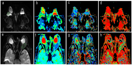
A total of 46 subjects with histologically confirmed orbital masses (34benign,12malignant) were enrolled in this retrospective study. All the participants underwent multi-b value diffusion imaging on a 3.0T whole body scanner (MR750, GE,USA) prior to a surgical therapy. 13 b values were acquired(0, 10, 20, 30, 50, 80, 100, 200, 300, 500, 600, 800 and 1000s/mm2). IVIM derived parameters including molecular diffusion coefficient(D), perfusion-related diffusion coefficient(D*), and perfusion fraction(f) were obtained using vendor supplied software. The ROIs were chosen to avoid contaminations from peripheral unintended tissues or inclusion of adjacent anatomical structures(Fig 1). Statistical analysis was performed using SPSS 22.0, A two-tailed P value of<0.05 were considered statistically significant. The diagnostic performances of D, D* and f were evaluated using the receiver operating characteristic (ROC) analysis.Results
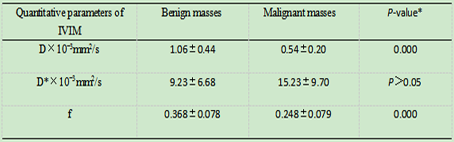
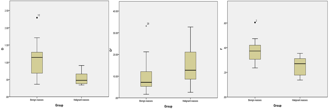
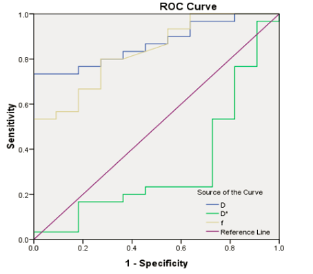
The IVIM parametric maps of a case of cavernous hemangioma and a case of orbital lymphoma are shown in Fig.1. Table 1 summarizes the IVIM-DWI parameters obtained from malignant and benign orbital masses. D and f values were significantly lower in malignant orbital masses than that in benign orbital masses (P<0.001). No significant difference was found in D* values in orbital masses(P>0.05). The box plots of IVIM parameters were shown in Fig. 2. ROC curve analysis showed that the area under the curve(AUC) of D and f values were 0.855 and 0.848, respectively in Fig. 3. The respective optimal cutoff values under the 95% CI according to best Youden index were: D=0.906×10⁻³mm2/s with a sensitivity of 67.6%, a specificity of 100%, and an accuracy of 76.1%, f=0.295 with a sensitivity of 79.4%, a specificity of 75%, and an accuracy of 78.2%.Discussion and conclusion
In this study, it was seen that benign and malignant masses in orbit feature distinctive D and f values derived from IVIM. IVIM DWI facilitated understanding of tumor tissue characteristics of perfusion and diffusion, and it could potentially serve as non-invasive predictors for the preoperative differentiation in orbital masses.Acknowledgements
We are gratefully thankful for the support and assistance from Hui Lin of the Advanced Application Team of GE Healthcare China.References
[1] Fatima Z, Ichikawa T, Ishigame K, et al. Orbital masses: the usefulness of diffusion-weighted imaging in lesion categorization. Clin Neuroradiol. 2014. 24(2): 129-34.
[2] Sepahdari AR, Aakalu VK, Setabutr P, Shiehmorteza M, Naheedy JH, Mafee MF. Indeterminate orbital masses: restricted diffusion at MR imaging with echo-planar diffusion-weighted imaging predicts malignancy. Radiology. 2010. 256(2): 554-64.
[3] Iima M, Le BD. Clinical Intravoxel Incoherent Motion and Diffusion MR Imaging: Past, Present, and Future. Radiology. 2016. 278(1): 13-32.
Figures