1840
Model-Based Joint Reconstruction for Multi b-Value Diffusion-Weighted Imaging1School of Biomedical Engineering, Guangdong Provincial Key Laborary of Medical Image Processing, Southern Medical University, Guangzhou, People's Republic of China, 2Philips Healthcare (Suzhou), Suzhou, People's Republic of China, 3Philips Healthcare, Guangzhou, People's Republic of China, 4Department of Biomedical Engineering, Zhejiang University, Hangzhou, People's Republic of China, 5laboratory of Biomedical Imaging and Signal Processing, The University of Hong Kong, Hong Kong SAR, People's Republic of China, 6Department of Electrical and Electronic Engineering,The University of Hong Kong, Hong Kong SAR, People's Republic of China, 7Neusoft Medical System, Shanghai, People's Republic of China
Synopsis
In current multi b-value DWI, each b-value image is usually reconstructed independently by using parallel MRI techniques. In this work, we propose a model-based joint reconstruction method for the reconstruction of under-sampled multi b-value DWI data. The proposed method can directly estimate quantitative parameters form k-space data, and exploit inter-image constraint to improve the quality of reconstructed image.
Purpose
Quantitative parameters measured from multi b-value diffusion weighted imaging (DWI) plays an important role in tissue characterization. In practice, multi b-value DWI requires to obtain images of multiple b-values and directions. In addition, high b-value DWI is usually acquired with multiple averages for sufficient signal to noise ratio. Thus, multi b-value DWI requires long acquisition time. Currently, each image in multi b-value DWI is usually reconstructed independently by using parallel MRI techniques that suffers from noise amplification due to the degraded matrix inversion condition. This work aims to develop a model-based joint reconstruction method for the reconstruction of under-sampled multi b-value DWI data.Method
Based on the assumption the signal decay in multi b-value DWI follows a mono-exponential function, the proposed joint reconstruction method can be formulated as:
$$\normalsize \min_{D, m_{0}} \sum_{j,k}\parallel d_{j,k}-RFS_{k}\phi _{j}m_{0}e^{-b_{j}D}\parallel_{2}+\lambda_{1}\parallel TV(m_{0}) \parallel_{1}+\lambda_{2}\parallel TV(D) \parallel_{1}$$
where m0 is the non-diffusion image, φj the phase of diffusion image with b-value of bj, D apparent diffusion coefficient (ADC), Sk the coil sensitivity of the k-th coil, F the fourier transform, R the acquisition pattern, dj,k the acquired k-space data and λ1 and λ2 are the regularization parameters. This joint reconstruction method directly estimates the ADC map and non-diffusion image from the k-space data. After that, the multi b-value DW images can be calculated from the estimated D and m0 maps by using the diffusion model. The above cost function is solved by the conjugate gradient algorithm1.
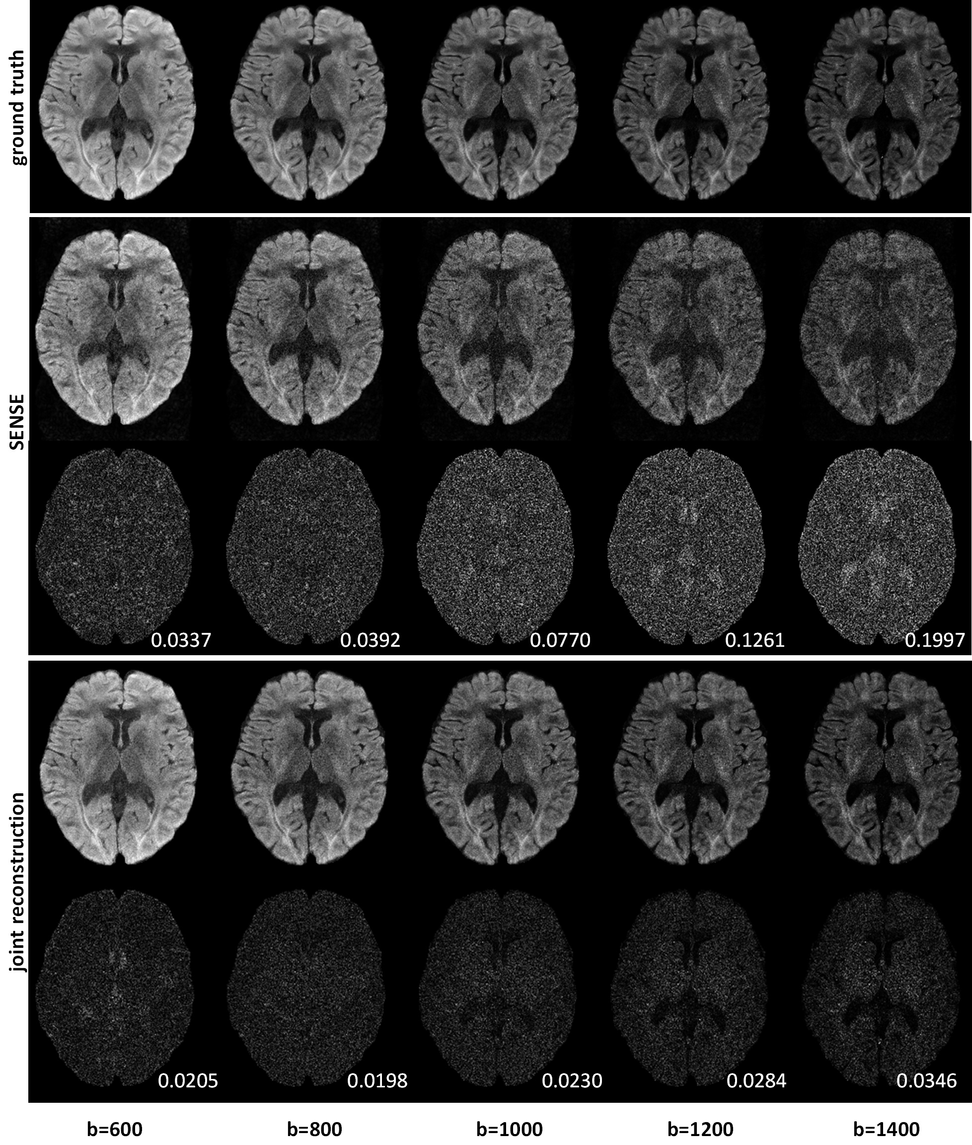
Simulation was performed to assess the performance of the proposed method. A set of non-diffusion and diffusion images reconstructed from 4-shot 8-channle EPI brain data set was used for data simulation. The ADC map was calculated and images at b-values of 600, 800, 1000, 1200 and 1400 s/mm2 were synthesized by using the mono-exponential diffusion model. Linear-phase ramps were then applied in the generated DW images to simulate motion-induced phase errors. Gaussian noise with a level of 1% was added to the complex images. The coil sensitivities were applied to these noise-corrupted images to simulate DW images acquired with an 8-channel coil. Fast Fourier transform was performed to transform the multiple coil images to multichannel k-space data. A quarter of the k-space data with uniform under-sampling was extracted for algorithm evaluation. The joint reconstruction was compared with independent SENSE2 reconstruction in terms of the estimated ADC map and multi b-value DW images.
Results
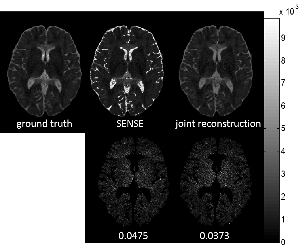
Figure 1 shows the ADC maps of ground truth, independent SENSE and the proposed method and the corresponding residual maps with respect to ground truth. The proposed method obtained more accurate ADC map than independent SENSE method. Figure 2 shows the multi-b DW images and residual maps between images reconstructed from different methods and ground truth. The proposed joint reconstruction method produced multi-b DW images with reduced noise compared to the independent SENSE method. Quantitatively, the proposed method yielded substantially decreased NRMSE values than the independent SENSE method.Discussion
The simulation results demonstrate the proposed model-based joint reconstruction method can produce improved multi-b DW images and ADC map. Though the proposed joint reconstruction method is implemented based on the mono-exponential diffusion model, other sophisticated models can be easily incorporated into the proposed framework.Conclusion
The proposed model-based multiple b-value joint reconstruction method can effectively improve the reconstruction quality of under-sampled multi b-value DWI data.Acknowledgements
No acknowledgement found.References
1. Lustig M, et al. Sparse MRI: the application of compressed sensing for rapid MR imaging. Magn Reson Med. 2007; 58:1182–1195.
2. Pruessmann KP, et al. SENSE: sensitivity encoding for fast MRI. Magn Reson Med. 1999; 42(5):952-962.
Figures