1839
Fractional motion related diffusion MRI in the detection of acute ischemic stroke1MR Research China, GE Healthcare, Beijing, People's Republic of China, 2Center for MR Research, Peking University, Beijing, People's Republic of China, 3Department of Radiology, Beijing Tiantan Hospital, Beijing, People's Republic of China
Synopsis
The use of FM model has been demonstrated in distinguishing low- and high-grade pediatric brain tumors. However, its feasibility in detecting acute stroke has not yet been investigated. In this work, FM model was applied in patients with acute ischemic stroke and compared with traditional ADC to investigate its clinical potential.
Purpose
Diffusion weighted imaging has become a pillar of magnetic resonance clinical imaging, especially in diagnosis of brain tumors and acute ischemia1. Comparing to the conventional Gaussian diffusion model, the fraction diffusion model (FM) has the potential to more accurately depict the diffusion process in biological tissues2. The use of FM model has been demonstrated in distinguishing low- and high-grade pediatric brain tumors3. However, its feasibility in detecting acute stroke has not yet been investigated. In this work, FM model was applied in patients with acute ischemic stroke and compared with traditional ADC to investigate its clinical potential.Methods
In this study, eight patients diagnosed with acute ischemic stroke were retrospectively extracted from the database of Beijing Tiantan Hospital. Specific DWI images of the whole brain were acquired within the first nine hours of stroke onset on a 3T GE Discovery MR750 MRI scanner. The diffusion gradients were applied successively in three orthogonal directions. In each direction, a total of 18 non-zero b-values were applied, ranging from 150 to 3500 s/mm2 with varying both diffusion gradient amplitudes and separation times. The obtained DWI images were first corrected for eddy currents distortions and head motions using FSL. FM related anomalous diffusion maps, α and H, were obtained by fitting DWI images with the FM model as shown in ref. 2. In addition, ADC value was computed using b = 1000 s/mm2, directionally averaged maps were then calculated to reduce the influence of anisotropy. The regions of interest of stroke lesions were manually drawn by a radiologist based on ADC map; the mirror regions of the lesion ROIs were used as healthy tissue for comparison. Paired t test was performed between the metrics of the lesion ROIs and the healthy tissue.Results
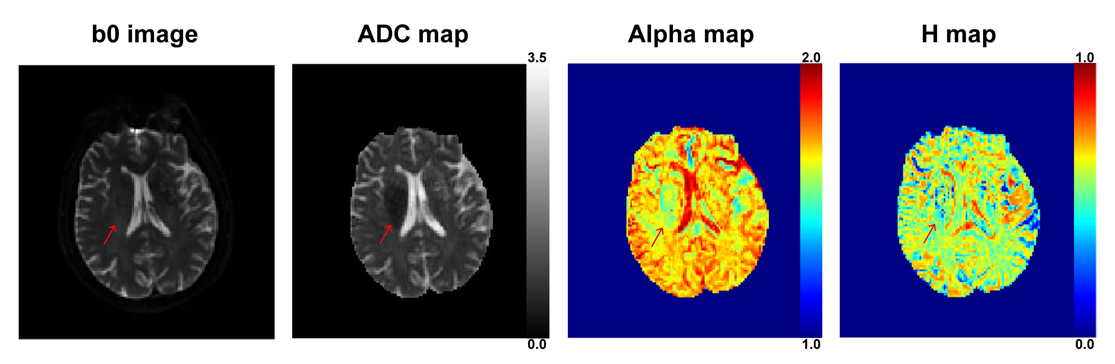
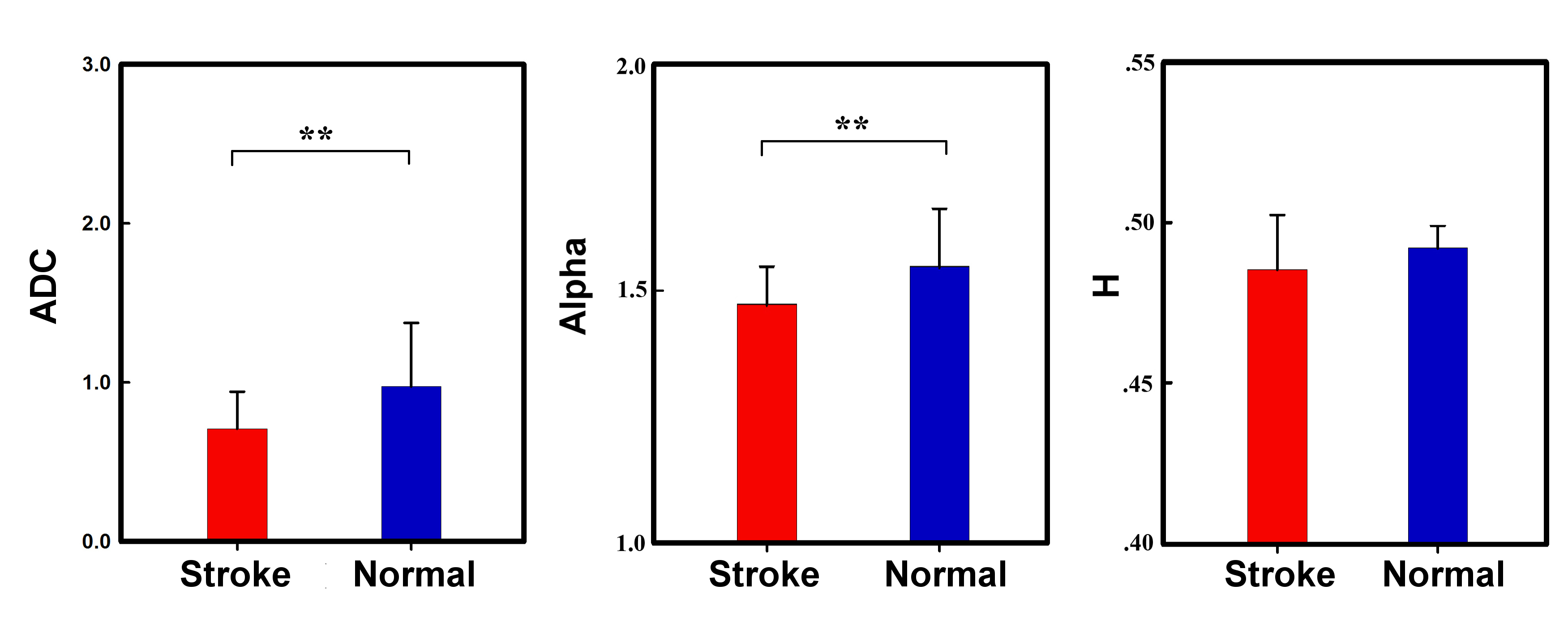
T2-weighted b0 image, ADC, α and H maps of a typical subject with acute stroke are shown in Fig. 1, where the arrows pointed the stroke lesion region. Lowered ADC value was observed in the lesion region as compared to the surrounding normal tissue. On the contrary, no significant signal change was seen in T2 weighted image. α and H maps also showed decreased value in the lesion region as compared to the normal tissues. The results of paired t test are shown in Fig. 2. It is seen that both ADC and α parameters showed statistically significant difference between stroke regions and normal tissues; although H featured decreased values such difference was not statistically significant.Discussion and Conclusion
In this feasibility study, the use of FM model in detecting acute ischemia stroke was investigated. Similar to conventional ADC, the FM model may be used for identifying stroke lesions surrounded by healthy tissues. The parameter α determines the distribution of microscopic jump length, and the parameter H governs the similarity of particles’ trajectories. Based on the results, the α value was more sensitive to stroke regions. The observed results indicate that the microscopic diffusion in the lesion region may further derivate away from Gaussian distribution as compared to normal tissues. Considering that the underlying principle of reduced diffusivity in stroke remains unknown4, the use of non-Gaussian model may further illuminate the underlying pathological cause. Further study with a larger cohort is needed in revealing the clinical value of FM model in acute stroke.Acknowledgements
No acknowledgement found.References
[1] Le Bihan, Denis, and Heidi Johansen-Berg. Diffusion MRI at 25: exploring brain tissue structure and function. Neuroimage 61.2 (2012): 324-341.
[2] Fan, Yang, and Jia-Hong Gao. Fractional motion model for characterization of anomalous diffusion from NMR signals. Physical Review E 92.1 (2015): 012707.
[3] Karaman, M. Muge, et al. A fractional motion diffusion model for grading pediatric brain tumors. NeuroImage: Clinical 12 (2016): 707-714.APA
[4] Le Bihan, Denis, and Mami Iima. Diffusion magnetic resonance imaging: what water tells us about biological tissues. PLoS Biol 13.7 (2015): e1002203.
Figures