1836
Diffusion weighted imaging in thyroid nodule: comparison of readout-segmented EPI and single-shot EPI Techniques1Radiology, Changhai Hospital of Shanghai, Shanghai, People's Republic of China, 2Application development, Siemens Shenzhen Magnetic Resonace Ltd, Shenzhen, People's Republic of China
Synopsis
Diffusion weighted imaging showed the potential to evaluate thyroid disease. This study aimed to evaluate whether readout-segmented EPI (RS-EPI) can provide better image quality in imaging thyroid gland in comparison with single-shot EPI (SS-EPI), and to compare ADC values, acquired from RS-EPI with those of SS-EPI. Sixteen patients were examined using both techniques. There were significant differences in susceptibility, motion artifacts, except for detectability of thyroid nodules and ADC measurements between RS-EPI and SS-EPI. The present study found that the RS-EPI technique provides significant image quality improvement compared with SS-EPI in imaging thyroid gland at 3 Tesla.
Purpose
Thyroid nodule is a common disorder of the thyroid gland [1]. Several studies have been reported that apparent diffusion coefficient (ADC) values can discriminate benign and malignant thyroid nodules [2-4]. Single-shot echo planar imaging (SS-EPI) is the most frequently used technique because of its relative insensitive to motion-induced phase errors, high signal-to-noise ratio and short acquisition times [5]. However, SS-EPI images are characterized by blurring along the phase encoding direction due to T2* decay and are sensitive to off-resonance effects [6], especially in the neck area. Recently, a novel multi-shot technique, called readout-segmented echo planar imaging (RS-EPI), has been proposed to improve the spatial resolution , decrease the susceptibility based image distortion and T2* blurring and a robust correction for motion-induced phase artifact [7]. We hypothesized and believed that the RS-EPI could have a promising application for diffusion weighted imaging (DWI) in thyroid gland, which is complex and prone to severe artefacts. Therefore, the aims of the present study are twofold, to evaluate whether RS-EPI can provide better image quality in imaging thyroid gland in comparison with SS-EPI, and to compare ADC values, acquired from RS-EPI with those of SS-EPI.Materials and Methods
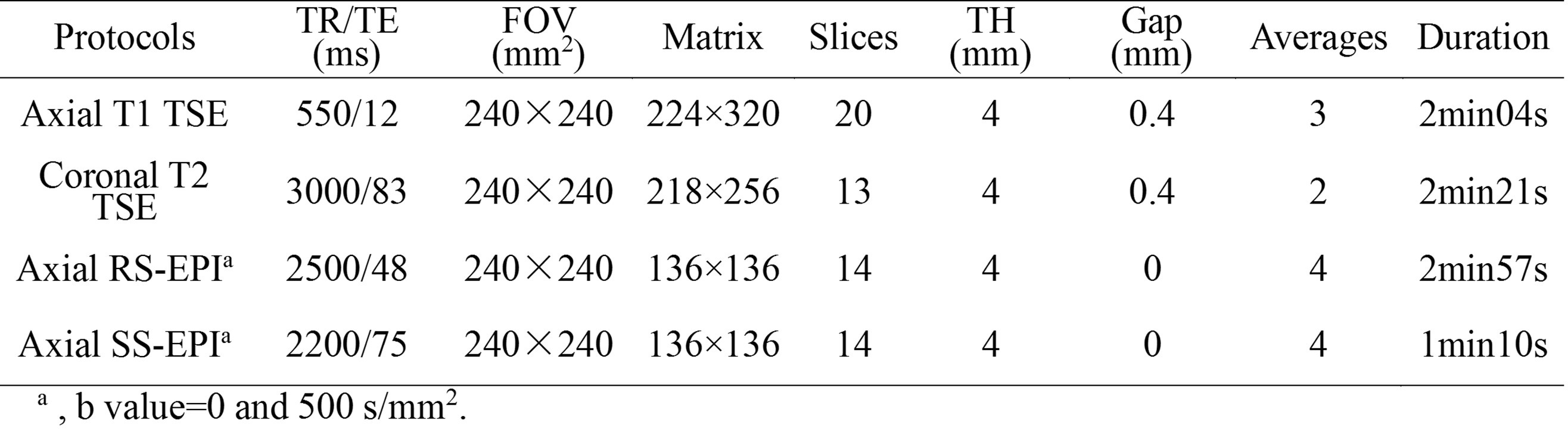
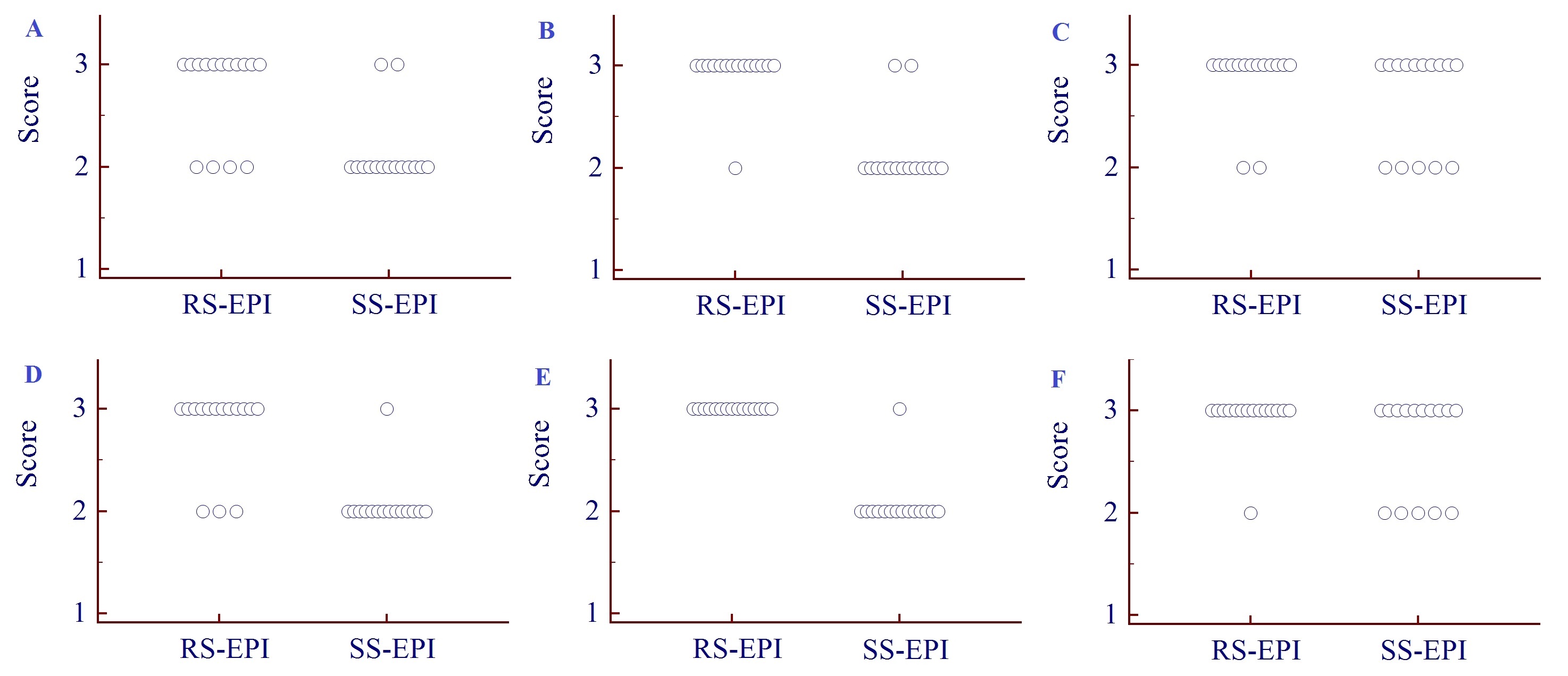
Subjects Sixteen patients (age, 45.8±16.8 years; male/female, 3/13) with thyroid disease were enrolled in this prospective study, which was approved by the local institutional review board and written informed consent was obtained from each patient. MRI protocols Magnetic resonance imaging was performed on a 3T MR system (Skyra, Siemens medical solution, Germany) using a standard 20-channel phased-array head/neck coil. In all patients, the examination protocols of thyroid gland included an axial T1-weighted turbo spin-echo (T1W TSE), a coronal T2-weighted TSE and two axial DWI with RS-EPI and SS-EPI techniques. T1W and T2W images were used for detecting the thyroid nodules and positioning the DWI sequences. The parameters of these protocols were summarized in Table 1. The total scan time of each subject was approximately 10 minutes. Image analysis RS-EPI and SS-EPI DWI images were evaluated by two independent observers (with 4 and 6 years of experience, respectively) for identification of susceptibility, motion artifacts (1, marked; 3, no artifact) and detectability of thyroid nodules (1, poor; 3, good) using quantitative scores. ADC values of thyroid nodules were measured and compared. The regions of interest (ROI) of thyroid nodules were manually outlined on ADC maps with the sizes of ROIs ranged from 1.05 mm2 to 4.2 mm2. Statistical analysis Significant differences between RS-EPI and SS-EPI for visual scores, and ADC value were assessed by using Wilcoxon signed rank tests.Results
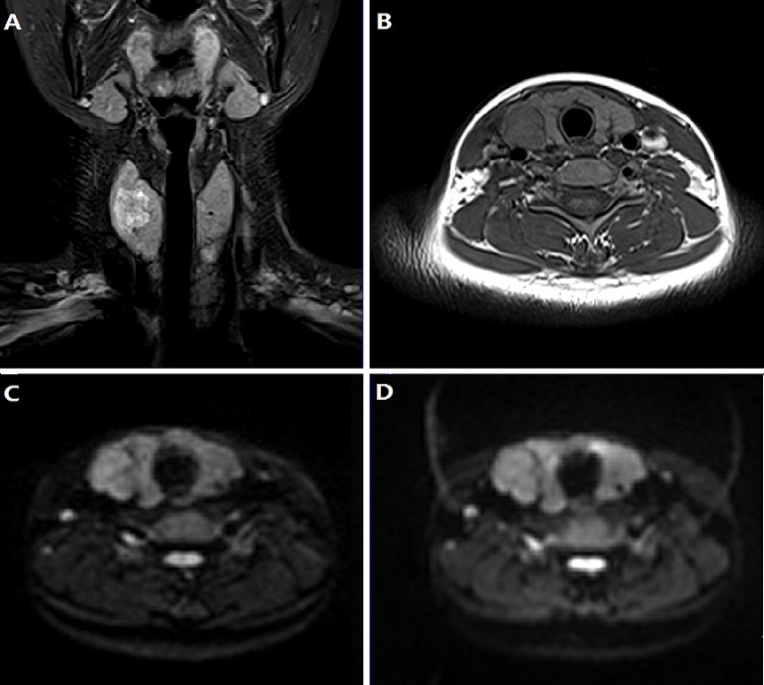
One patient was excluded in the final image analysis due to poor image quality and uncooperative examination. The statistical results of quantitative scores in assessing thyroid nodules were summarized in Table 2. There were significant differences in susceptibility (Figure 1A, D, P < 0.05), motion artifacts (Figure 1B, E, P < 0.05), except for detectability of thyroid nodules (Figure 1C, F, P > 0.05). RS-EPI technique is superior to SS-EPI technique in image quality (Figure 2). No significant difference was observed for ADC measurements of thyroid nodules between RS-EPI and SS-EPI techniques (1.629±0.539×10⁻³ mm/s and 1.623±0.596×10⁻³ mm/s, respectively, P=0.8469).Discussion and Conclusion
In this study, we demonstrated significantly higher image quality of thyroid DWI using RS-EPI than that using the conventional SS-EPI technique, with no significant difference in ADC values, which indicated that RS-EPI DWI is an effective method for reducing image distortion, artefacts and improving image quality in thyroid gland. The distortion in DWI was significantly reduced with RS-EPI. The RS-EPI technique provides significant image quality improvement compared with SS-EPI at 3 Tesla, and can potentially offer high image quality in imaging thyroid gland.Acknowledgements
No acknowledgement found.References
[1] Erdem G, Erdem T, Muammer H, et al. Diffusion-weighted images differentiate benign from malignant thyroid nodules. J Magn Reson Imaging. 2010;31(1):94–100.
[2] Noda Y, Kanematsu M, Goshima S, et al. MRI of the thyroid for differential diagnosis of benign thyroid nodules and papillary carcinomas. AJR Am J Roentgenol. 2015;204(3):W332-335.
[3] Ilica AT, Artas H, Ayan A, et al. Initial experience of 3 tesla apparent diffusion coefficient values in differentiating benign and malignant thyroid nodules. J Magn Reson Imaging. 2013;37(5):1077-1082.
[4] Shi HF, Feng Q, Qiang JW, et al. Utility of diffusion-weighted imaging in differentiating malignant from benign thyroid nodules with magnetic resonance imaging and pathologic correlation. J Comput Assist Tomogr. 2013;37(4):505-510.
[5] Turner R, Lebihan D. Single-Shot Diffusion Imaging at 2.0 Tesla. J Magn Reson. 1990;86: 445–452.
[6] Taviani V, Nagala S, Priest AN, et al. 3T diffusion-weighted MRI of the thyroid gland with reduced distortion: preliminary results. Br J Radiol. 2013;86(1028):20130022.
[7] Porter DA, Heidemann RM. High resolution diffusion-weighted imaging using readout-segmented echo-planar imaging, parallel imaging and a two-dimensional navigator-based reacquisition. Magn Reson Med. 2009;62(2):468-475.
Figures