1829
Effect of gadolinium contrast agent on IVIM derived parameters of abdominal organs1Radiology, Changhai Hospital of Shanghai, Shanghai, People's Republic of China, 2MR Collaboration NE Asia, Siemens Healthcare, Shanghai, People's Republic of China
Synopsis
This study investigated potential effects of gadolinium contrast agent on the IVIM derived parameters such as Dfast (blood microcirculation), Dslow (pure extravascular water diffusion), f (perfusion fraction) and the commonly used DWI-derived ADC of abdominal organs. The result shows that gadolinium administration does not make statistically significant differences in Dslow, Dfast, f or ADC of the liver, spleen, or pancreas. In the kidney, however, ADC values are significantly lower with post-contrast than pre-contrast.
Background and Purpose
Diffusion-weighted magnetic resonance imaging (DWI) is a noninvasive technique exploring the microscopic mobility of water molecules in the tissues without contrast administration 1. Recent technique advancements allow DWI and apparent diffusion coefficient (ADC) measurements to be increasingly used in the evaluation of abdominal diseases 2-4. The effects of gadolinium contrast agent on DWI of the liver, spleen, pancreas and kidney have been reported. These reports, however, used only two or three b values to measure monoexponential ADC, which is influenced not only by the structures of the tissue, but also by the microcirculation of blood in the capillary network. Ideally, multiple-b-value DWI with intravoxel incoherent motion (IVIM) model should be set up for the separated estimation of tissue perfusion and diffusivity(5). The aim of this study was to investigate potential effects of gadolinium contrast agent on the IVIM derived parameters such as Dfast (blood microcirculation, perfusion), Dslow (pure extravascular water diffusion), f (perfusion fraction) and the commonly used DWI-derived ADC of the liver, spleen, pancreas and kidney.Methods
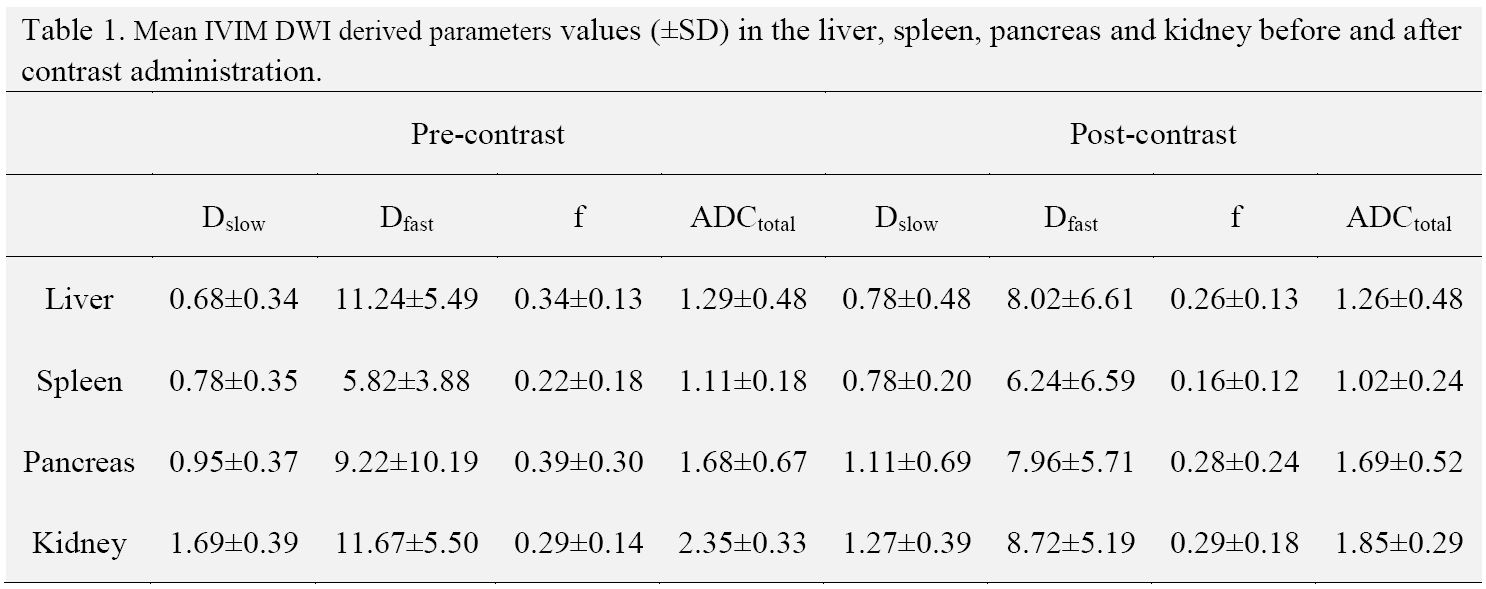
This study was approved by our institution review board. Informed consent was obtained from all subjects. 11 patients were recruited and underwent multiple b-value DWI (b values = 0, 100, 500, 800 s/mm2) before and after gadolinium agent was injected on a 3T whole body system (MAGNETOM Skyra, Siemens healthcare, Erlangen, Germany). ADC map was generated inline automatically by Syngo software, while IVIM derived parameters were calculated offline by using a prototype software called body diffusion toolbox. Diffusion parameters including ADC and biexponential IVIM parameters (Dslow, Dfast and f) were measured for the normal liver, spleen, pancreas and kidney using free hand regions of interest.Results
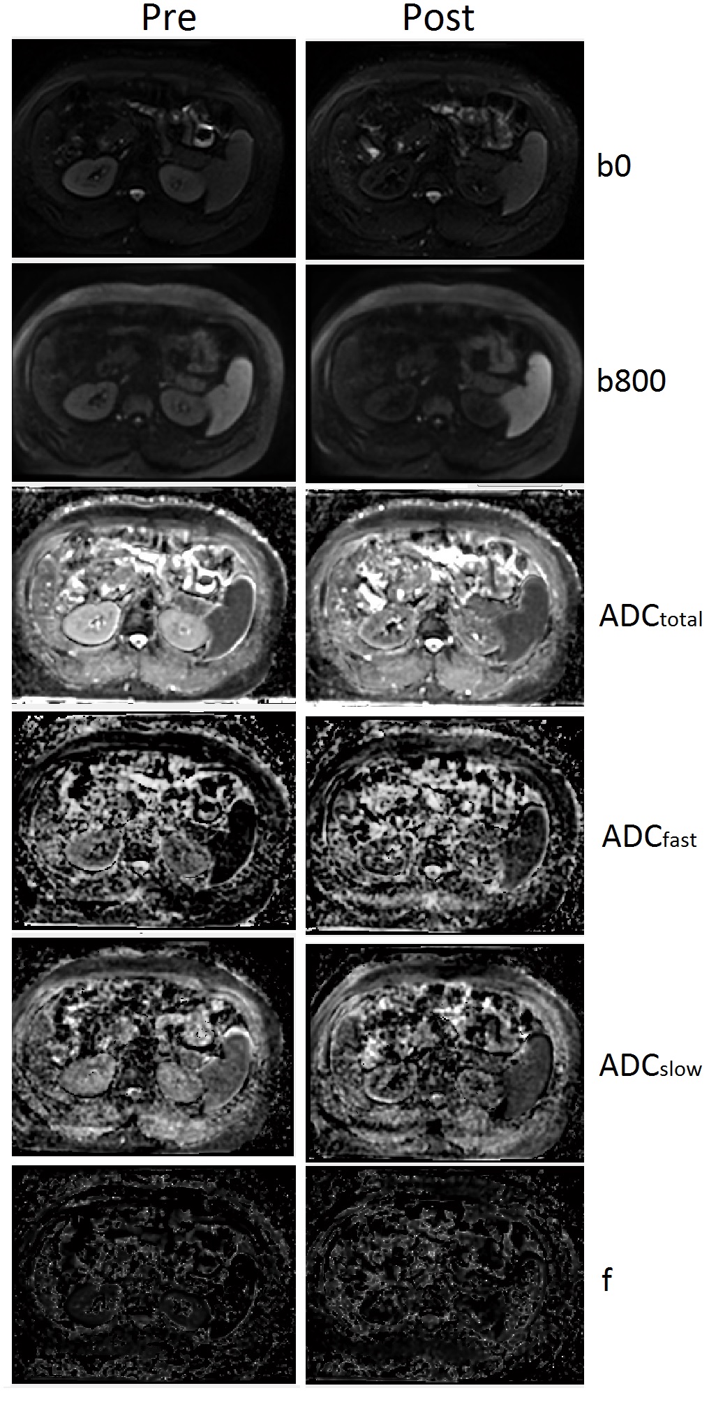
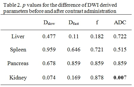
The typical ADC and IVIM parameters maps are demonstrated in Figure 1. Most parameters showed lower values on the liver, spleen, pancreas and kidney in post-contrast than in pre-contrast. Among them, ADC value is significant lower in post-contrast compared with the pre-contrast on kidney, with p < 0.05. The results were showed and summarized in Table 1 and Table 2.
Conclusions
Intravenous gadolinium administration does not make a statistically significant difference in the qualitative IVIM and DWI derived parameters including Dslow, Dfast, f or ADC of the liver, spleen, or pancreas. In the kidney, however, ADC values are significantly lower with post-contrast than pre-contrast.
Acknowledgements
This work was supported by the Natural Science Foundation of Shanghai (14ZR1408300); medical guidance project of Shanghai Municipal Science and Technology Commission (14411960100); the 1255 Academic Discipline Project of Shanghai Changhai Hospital (CH125520800); the Youth Scientific Research Funds of Shanghai Changhai Hosptial.References
1. Bammer R. Basic principles of diffusion-weighted imaging. Eur J Radiol. 2003 Mar;45(3):169-184.
2.Dale BM, Braithwaite AC, Boll DT, et al. Field strength and diffusion encoding technique affect the apparent diffusion coefficient measurements in diffusion-weighted imaging of the abdomen. Invest Radiol. 2010 Feb;45(2):104-108.
3.Mürtz P, Flacke S, Träber F, et al. Abdomen: diffusion-weighted MR imaging with pulse-triggered single-shot sequences. Radiology. 2002 Jul;224(1):258-264.
4.Thoeny HC, De Keyzer F. Extracranial applications of diffusion-weighted magnetic resonance imaging. Eur Radiol. 2007 Jun;17(6):1385-1393.
5.Le Bihan D, Breton E, Lallemand D, et al. MR imaging of intravoxel incoherent motions: application to diffusion and perfusion in neurologic disorders. Radiology. 1986 Nov;161(2):401-407.
Figures