1826
Optimal strategy for measuring intraventricular temperature using second-order motion compensation DWI1Department of Radiology, Tokai university hospital, Isehara, Japan, 2Division of Health Sciences, Graduate School of Medical Sciences, Kanazawa University, Kanazawa, Japan, 3Radiology, Tokai University School of Medicine, Isehara, Japan, 4Healthcare department, Philips Electronics Japan, LTD, Japan
Synopsis
A method for monitoring the intraventricular cerebrospinal fluid (CSF) temperature calculated from the DWI is affected by the CSF pulsation. Moreover, DWI should be obtained by optimal b value according to the diffusion coefficient of the measuring tissues. We investigated the second-order motion compensation DWI (2nd-MC DWI) to the determination of the intraventricular temperature to improve that accuracy with optimal b value. In the case of using the optimal b value based on the literature (ie., approximately 400 s/mm2), the intraventricular temperature can be more accurately estimated with 2nd-MC DWI than conventional
INTRODUCTION
Intraventricular temperature can be estimated by diffusion weighted imaging (DWI) using the diffusion coefficient of cerebrospinal fluid (CSF) that reflects the temperature of water molecules1. However, this method may be affected by the CSF pulsation, which causes an error in the estimation of the temperature in CSF. Because DWI should be obtained by optimal b value according to the diffusion coefficient of the measuring tissues2, low b value should be used for the region such as CSF space where the diffusion coefficient is higher than the other brain tissues. However, at low b value DWI, the influence of CSF flow may significantly affect intraventricular temperature estimation. Here, we applied the second-order motion compensation DWI (2nd-MC DWI) for accurate determination of the intraventricular temperature.METHODS
Eight healthy volunteers were examined at a 3.0 T MR system (Achieva R2; Philips Healthcare, Best, the Netherlands). DWI of the lateral ventricle (LV) and third ventricle (TV) were obtained. We assessed the accuracies of the measurement of the intraventricular temperature between no motion compensation DWI (no-MC DWI) and 2nd-MC DWI (Fig.1). Assuming that the diffusion coefficient of free water on 37 degrees C is 3.0 × 10⁻³ mm2/s 3, the optimal b value should be 426 × 10-6 s/mm2, based on the literature2. Therefore, five b values (i.e., 200, 400, 600, 800 and 1000 s/mm2) were applied, including possible optimal b value and b = 1000 commonly used on the brain DWI. We performed two-dimensional single-shot echo-planar imaging (EPI) DWI with the following parameters: TR, 6000 ms; TE, 90 ms; flip angle, 90 degrees; field of view, 220 × 220 mm2; matrix,192 × 192; slice thickness, 3.0 mm, number of slices, 30; EPI factor, 35, and sensitivity encoding factor, 3. To evaluate the signal change in CSF depending on each b value, the ratios of signal intensity on DWI with each b value to that on no diffusion weighting (Sb/S0) were calculated in LV and TV. Then, we created intraventricular temperature map calculated using diffusion coefficient from the DWI images1, and the intraventricular temperature in each ventricle was analyzed. The differences of the intraventricular temperature for each b value were assessed using Friedman test and a post-hoc test on each DWI technique.RESULTS and DISCUSSION
Examples of intraventricular temperature maps in each b value are shown in Fig.2. The ratio of signal intensity for no-MC DWI in TV was clearly demonstrated to be lower at lower b values than those for 2nd-MC DWI (Fig.3). This result suggests that no-MC DWI is more strongly affected by CSF flow and pulsation, particularly at the low b value. In the LV for each DWI technique, the intraventricular temperature was not significantly different among five b values (Table 1). The intraventricular temperature in the TV was significantly higher with no-MC DWI with b = 200 and 400 than with b = 1000; whereas only 2nd-MC DWI with b = 200 was significantly higher than that with b = 1000. Because the mean normal brain temperature has been reported as 38.1 or 36.5 degrees C in the previous studies4,5, the intraventricular temperature can be more accurately estimated with 2nd-MC DWI than no-MC DWI, particularly in TV. In addition, The estimate of temperatures at LV, where little pulsation was considered, were close to the normal brain temperatures in all b values on 2nd-MC DWI, but those on no-MC DWI showed over 40 degrees C at b value = 200 and 400. Therefore, 2nd-MC DWI can provide robust performance for analyzing the intraventricular temperature on the optimal b value.CONCLUSION
The 2nd-MC DWI may be the optimal strategy for measuring accurately the intraventricular temperature.Acknowledgements
No acknowledgement found.References
1. Delannoy J, et al. Magn Reson Med. 1991; 19: 333-9.
2. Saritas EU, et al. IEEE Trans Med Imaging. 2011; 30: 424-37.
3. Tofts PS, et al. Magn Reson Med. 2008; 59: 190-5.
4. Childs C et al. Magn Reson Med. 2007; 57: 59-66.
5. Covaciu L et al. J Magn Reson Imaging. 2010; 31: 807-14.
Figures
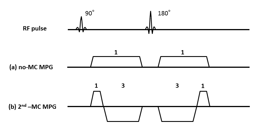

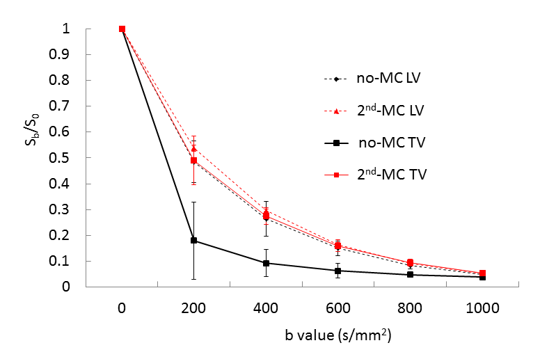
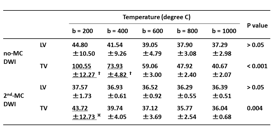
Table 1. Mean intraventricular temperature in each ventricle for the two DWI techniques.
Daggers indicate significant differences, compared with the temperature on no-MC DWI at b = 1000 s/mm2 (P < 0.05). Asterisk indicates significant differences, compared with the temperature on 2nd-MC DWI at b = 1000 s/mm2 (P < 0.05).