1819
High-Resolution Intravoxel Incoherent Motion (IVIM) with Generalized SLIce Dithered Enhanced Resolution Simultaneous MultiSlice (gSlider-SMS): Effect on CSF Partial Volume Contamination1Department of Medical Imaging, University of Toronto, Toronto, ON, Canada, 2Athinoula A. Martinos Center for Biomedical Imaging, Massachusetts General Hospital, Charlestown, MA, United States
Synopsis
In this work, we introduce a high-resolution acquisition strategy for IVIM brain imaging using generalized Slider Simultaneous MultiSlice (gSlider-SMS) diffusion MRI. An SNR efficient multi b-value 10 simultaneous slice acquisition was used to obtain IVIM parameter maps with submillimeter isotropic resolution. Compared to a conventional acquisition (2 mm isotropic), high resolution IVIM provided improved delineation of the cortex and reduced partial volume contamination with CSF. Cortical perfusion measurements using the standard acquisition were falsely elevated by approximately 40% compared to gSlider-SMS IVIM. High resolution IVIM may provide more reliable perfusion information for evaluation of cortically based pathology.
Introduction
Despite a recent resurgence, brain imaging with IVIM remains technically challenging in part due to substantial CSF partial volume effects. Bulk CSF flow results in signal loss at low b-values (<200 s/mm2), the same regime sensitive to incoherent motion of intravascular water. CSF within a voxel thus falsely elevates the observed intravascular compartment fraction (perfusion fraction)1. This is particularly problematic for evaluation of the cortex and cortically based pathology. CSF contamination can be reduced using a 180° inversion pulse1 or T2-preparation module2, however these strategies result in signal loss due to incomplete recovery of brain magnetization at the time of excitation.
Here, we propose an alternate strategy to mitigate CSF contamination, by implementing IVIM using an SNR-efficient high-resolution diffusion MRI acquisition. Generalized SLIce Dithered Enhanced Resolution Simultaneous Multi-Slice (gSlider-SMS) combines blipped-CAIPI Simultaneous Multi-Slice excitation3 with RF encoding and super-resolution reconstruction over the slice encoded direction (gSlider) to achieve wholebrain diffusion MRI with submillimeter isotropic resolution4. We hypothesized that gSlider-SMS IVIM would result in reduced CSF partial volume contamination and improved delineation of the cortical GM-CSF interface.
Methods
Imaging was performed on the 3T Skyra Connectom Scanner (maximum gradient strength 300 mT/m) using a custom 64-channel receiver coil. A volunteer with no history of neurological disease underwent conventional and high-resolution (gSlider-SMS) diffusion MRI with b-values of 0, 250, 500, 750, and 1000 s/mm2. For high-resolution IVIM, 10 simultaneous slice sagittal acquisition was performed (gSlider×MB = 5×2) with zoomed imaging (ZOOPPA)5 (RZOOM=1.67) and iPAT=2 for a total in-plane acceleration of 3.33x, 190 slices providing whole-brain coverage with 64 directions / b-value, nominal voxel size 0.86×0.86×0.86 mm, gSlider slice-encoding thickness 4.3 mm with final nominal slice resolution of 4.3 mm / 5 = 0.86 mm, TR/TE = 4200/70 ms, effective echo spacing 0.31 ms, and partial Fourier 6/8 with POCS reconstruction to fill the missing k-space data. Conventional IVIM was performed using SMS diffusion-weighted EPI with nominal voxel size 2.0×2.0×2.0 mm, 6 directions / b-value, iPAT=2, MB=2, and TR/TE = 3000/55 ms. T1-weighted MP-RAGE was obtained for anatomical coregistration and segmentation using a 1.0 mm nominal isotropic voxel size.
Motion and eddy current correction was performed in FSL (http://fsl.fmrib.ox.ac.uk). Diffusion-weighted trace images were calculated for each b-value. The IVIM perfusion fraction (f) and diffusion coefficient (D) were estimated for each voxel using linear fitting of the log normalized signal curves for b > 200,6 a procedure which has been validated against nonlinear fitting of the full biexponential model.7 FreeSurfer (http://freesurfer.net) was used to coregister diffusion datasets to the MP-RAGE acquisition and segment the pial surface and cortical GM-WM interface. Regions of interest for WM and cortical GM were automatically generated in FreeSurfer, and the mean values of f and D were calculated for each ROI.
Results
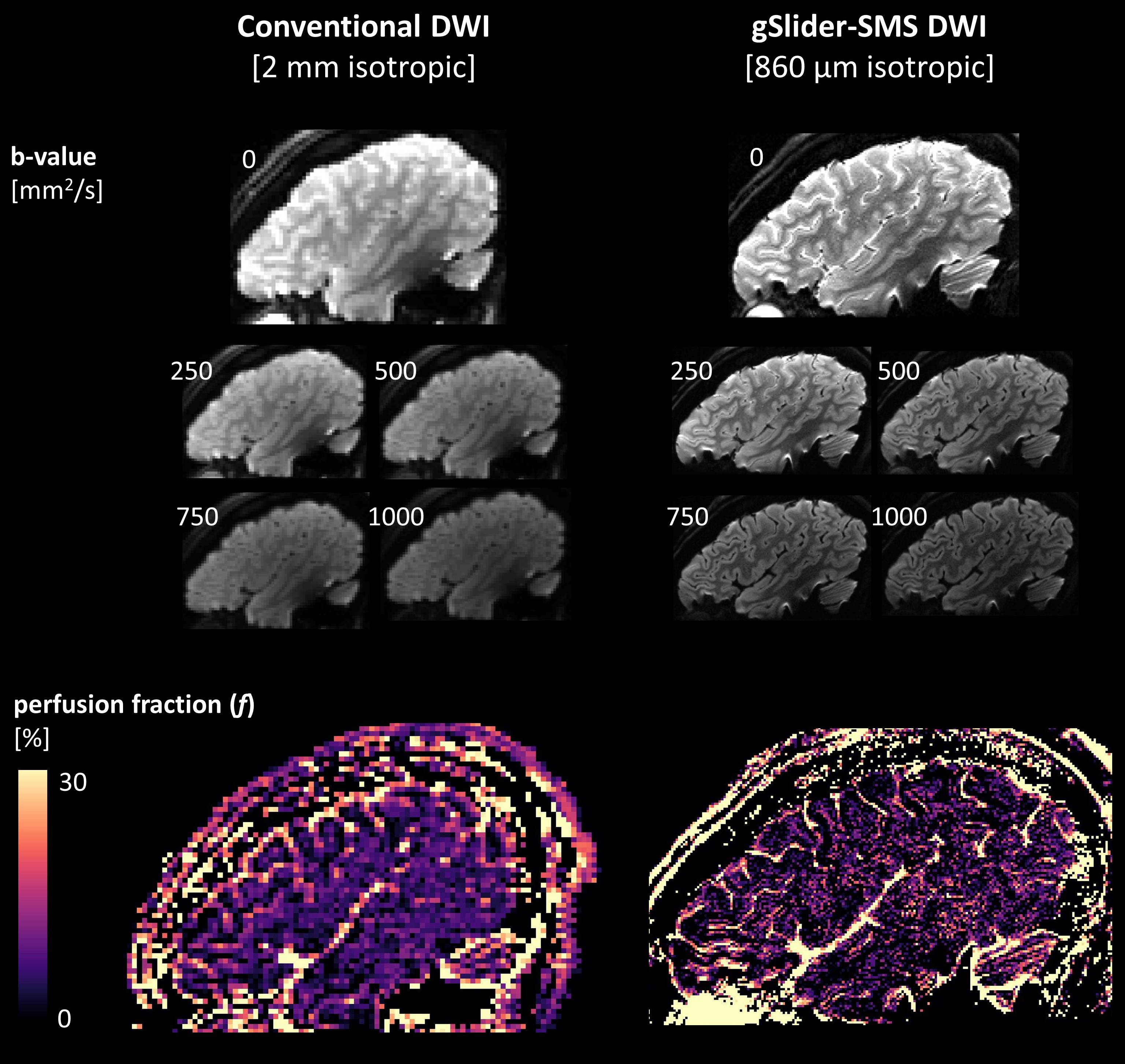
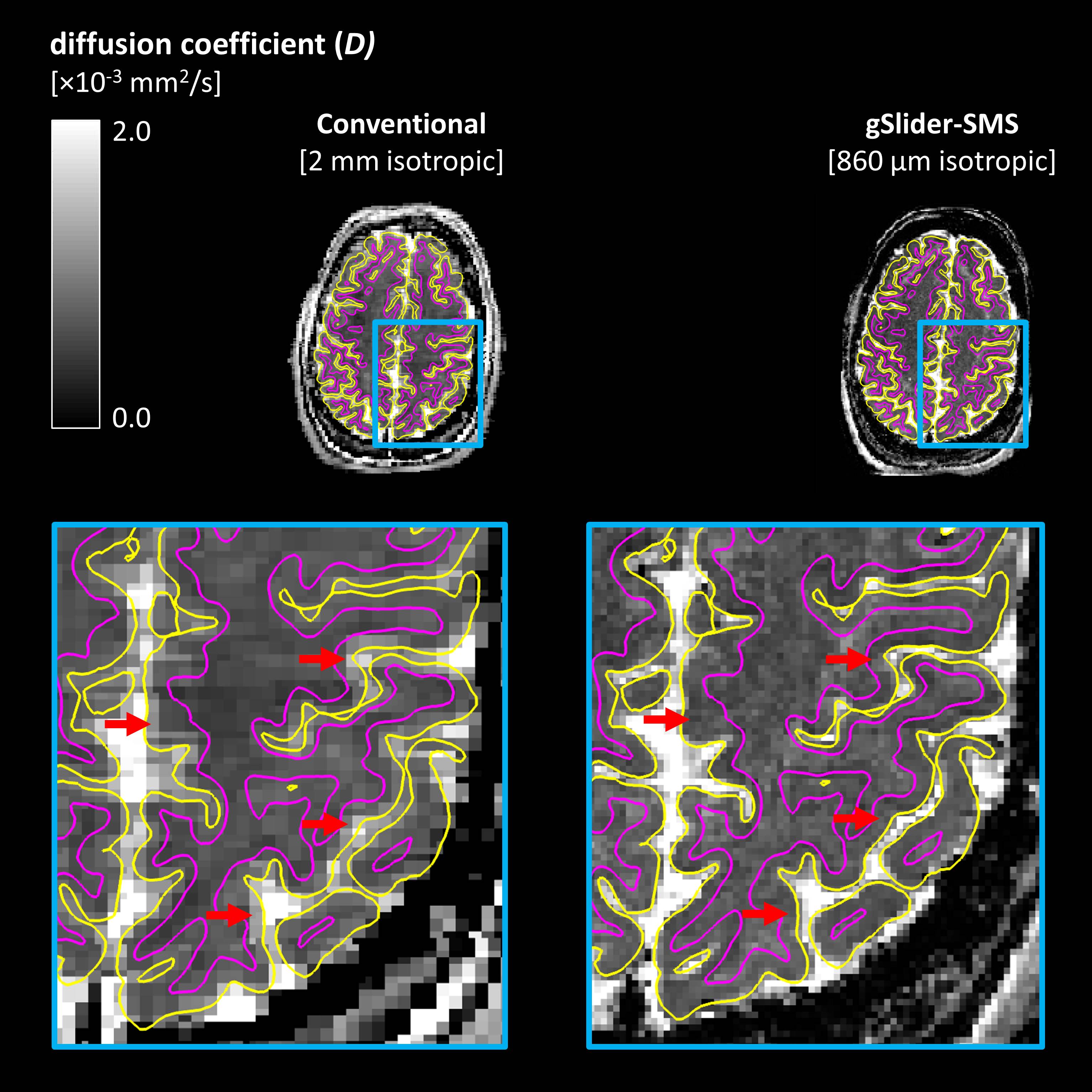
Representative diffusion-weighted images and perfusion fraction (f) maps for conventional and high-resolution (gSlider-SMS) IVIM are shown in Figure 1. Partial volume averaging is seen as intermediate valued (“pink”) voxels at the interface between CSF and cortical GM, particularly evident on the conventional acquisition. Figure 2 illustrates anatomical segmentation of the pial surface and WM-GM interface obtained using FreeSurfer, overlaid on coregistered maps of the diffusion coefficient (D). The high-resolution acquisition provided sharper delineation of the cortical mantle, more closely matching the automatically segmented cortical contours. Mean parameter values at 2 mm resolution were fGM=6.6%, fWM=5.1%, DGM=0.83×10⁻³ mm2/s, and DWM=0.75×10⁻³ mm2/s. Mean parameter values at 0.85 mm resolution were fGM=4.7%, fWM=4.3%, DGM=0.79×10⁻³ mm2/s, and DWM=0.80×10⁻³ mm2/s.Discussion and Conclusions
IVIM was successfully implemented at submillimeter isotropic resolution using gSlider-SMS diffusion MRI. In the conventional acquisition, elevated perfusion fraction and diffusion coefficient were observed in cortical GM relative to WM, consistent with CSF partial volume averaging. In contrast, high-resolution IVIM provided similar values of f and D for WM and cortical GM, as expected in the absence of significant CSF contamination. We observed a 40% increase in cortical perfusion fraction at 2 mm compared to 0.86 mm resolution, attributable to CSF partial volume effects. The ability to obtain IVIM perfusion measurements with high anatomic specificity may be especially valuable for evaluation of cortically-based pathology, and in future study of the physiologic basis of the IVIM parameters themselves. Limitations of this study include the small sample size (N=1) and simplified IVIM fitting procedure which precludes estimation of the pseudodiffusion coefficient, D*. Future work will apply gSlider-SMS IVIM to a larger cohort of healthy volunteers and patients with primary brain tumors (where IVIM has shown early clinical promise)8, and will extend the range of b-values to enable estimation of the full biexponential model parameters.Acknowledgements
This work was supported by the following NIH grants: P41EB015896, R01EB020613, U01MH093765, and instrumentation grants: S10-RR023401, S10-RR023043, and S10-RR019307.References
1. Kwong KK, McKinstry RC, Chien D, Crawley AP, Pearlman JD, Rosen BR. CSF-suppressed quantitative single-shot diffusion imaging. Magn Reson Med 1991;21(1):157-163.
2. Federau C, O'Brien K. Increased brain perfusion contrast with T2-prepared intravoxel incoherent motion (T2prep IVIM) MRI. NMR Biomed 2015;28(1):9-16.
3. Setsompop K, Gagoski BA, Polimeni JR, Witzel T, Wedeen VJ, Wald LL. Blipped-controlled aliasing in parallel imaging for simultaneous multislice echo planar imaging with reduced g-factor penalty. Magn Reson Med 2012;67(5):1210-1224.
4. Setsompop K, Stockmann J, Fan Q, Witzel T, Wald LL. Generalized SLIce Dithered Enhanced Resolution Simultaneous MultiSlice (gSlider-SMS) to increase volume encoding, SNR and partition profile fidelity in high-resolution diffusion imaging. Proc ISMRM 2016.
5. Heidemann RM, Anwander A, Feiweier T, Knösche TR, Turner R. k-space and q-space: combining ultra-high spatial and angular resolution in diffusion imaging using ZOOPPA at 7 T. Neuroimage 2012;60(2):967-978.
6. Le Bihan D, Breton E, Lallemand D, Aubin M, Vignaud J, Laval-Jeantet M. Separation of diffusion and perfusion in intravoxel incoherent motion MR imaging. Radiology 1988;168(2):497-505.
7. Conklin J, Heyn C, Roux M, Cerny M, Wintermark M, Federau C. A simplified model for intravoxel incoherent motion perfusion imaging of the brain. Am Journal Neuroradiol 2016; Epub ahead of print.
8. Puig J, Sánchez-González J, Blasco G, Daunis-i-Estadella P, Federau C, Alberich-Bayarri Á, Biarnes C, Nael K, Essig M, Jain R, Wintermark M, Pedraza S. Intravoxel Incoherent Motion Metrics as Potential Biomarkers for Survival in Glioblastoma. PLoS ONE 2016;11(7):e0158887.
Figures