1817
Potential value of turbo-spin echo diffusion-weighted imaging in nasopharynx: primary study for differential diagnosis between recurrent nasopharyngeal carcinoma and post-chemoradiation fibrosis1Radiology, Guangdong General hospital, Guangzhou, People's Republic of China
Synopsis
In our study, we found significantly higher image quality, lesion conspicuity and less distortion of TSE-DWI based on Alsop method compared with SS-EPI according to image quality scores. Frustratingly, the SNR for TSE-DWI was lower than for SS-EPI and the result was consistent with other non EPI-DWI sequences in previous studies. Clinically, the low SNR of TSE-DWI remains a major concern. May be it is contributed to the long reception time and higher actual resolution of TSE images compared with SS-EPI and the more efficient k-space coverage of SS-EPI. The CNR of nasopharynx lesions on DW images was significantly better for TSE-DWI imaging than for SS-EPI imaging, which enables a better visual discrimination of nasopharynx lesions with TSE-DWI imaging. In conclusion, TSE-DWI with fewer artifacts and much higher resolutions, which was near impossible before, will make up for the slightly poorer SNR. The ADC values of the brainstem, which was less affected by the susceptibility artifacts and ghosts, showed no significant differences between the two DWI techniques. However, the ADC values of the lesions on TSE were significantly different than those on SS-EPI. This result is obtained coincide with the previous result.These differences may be primarily attributed to susceptibility artifacts and ghost that also resulted in inhomogeneous ADC maps because nasopharyngeal DWI is vulnerable to these artifacts. Therefore, the ADC measurements from TSE-DWI might be more accurate than those from SS-EPI.
Introduction
To assess the diagnostic performance of the turbo-spin echo (TSE) diffusion-weighted imaging (DWI) in discriminating recurrent nasopharyngeal carcinoma (rNPC) from post- chemoradiation fibrosis.Methods
Twenty-seven (44%) patients with newly diagnosed rNPC and thirty-five (56%) patients with biopsy-proven post-chemoradiation fibrosis were recruited into tumor and fibrosis groups, respectively. All patients underwent two sets of nasopharyngeal DWI scan including TSE-DWI and single-shot echo-planar imaging (SS-EPI) DWI at a (Ingenia; Philips Healthcare) 3.0 T MR unit, and image quality was assessed by two experienced radiologists. DWI was performed with two b values (0 and 800 s/mm2). Statistical analyses were performed using the SPSS 20.0. Statistical significance was defined as P < 0.05. Paired Wilcoxon signed rank tests were used to assess the two radiologists’ ratings of the DWI images acquired using the two methods. Paired tests were used to compare the differences between the mean ADC values, and the SNRs and CNRs of the TSE and SS-EPI images. We used the mean ADC of both readers to optimize the ADC threshold for our data so as to reach the highest diagnostic accuracy based on receiver operating characteristic (ROC) analysis, and the corresponding diagnostic sensitivity, specificity were determined.Results
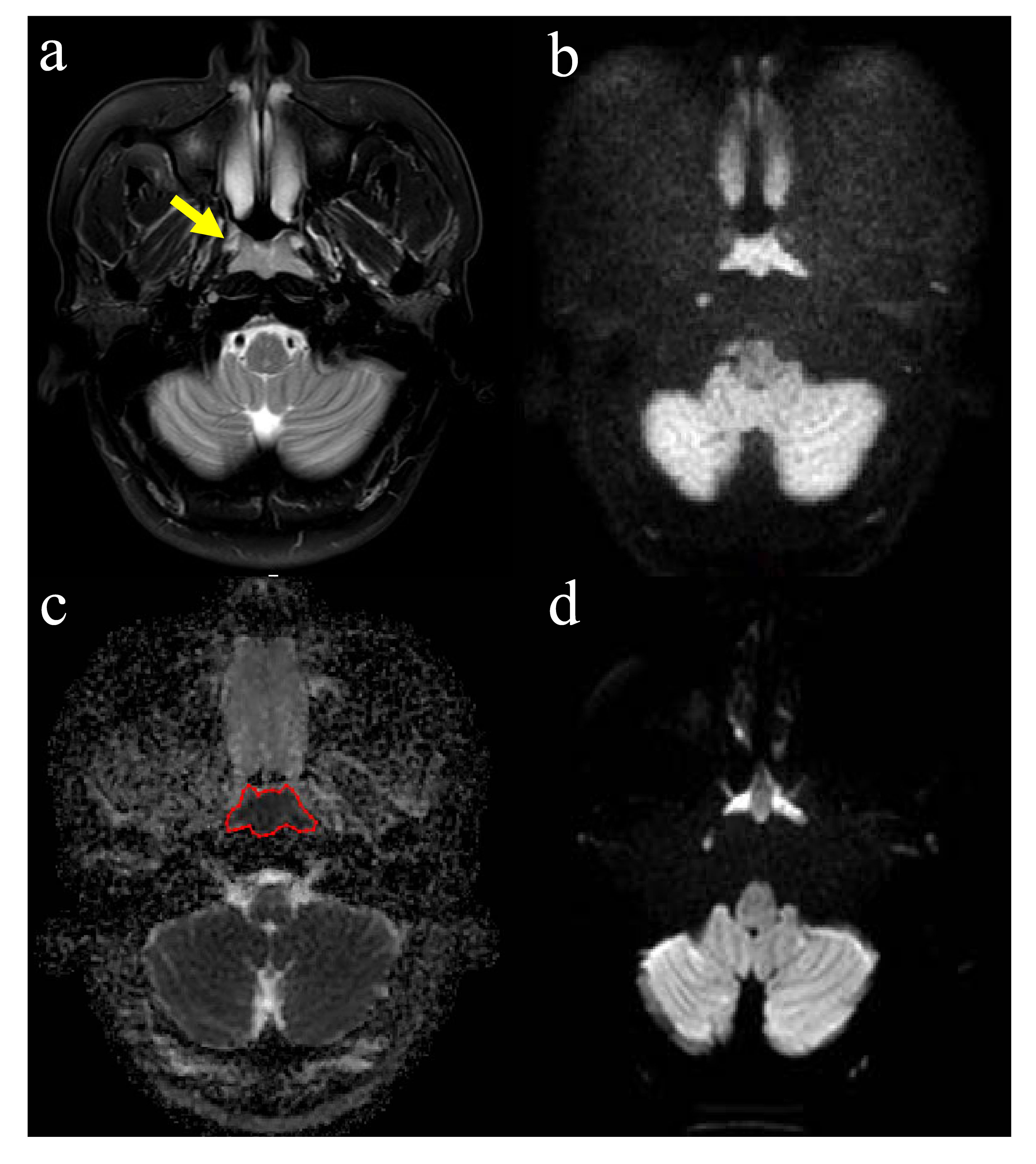
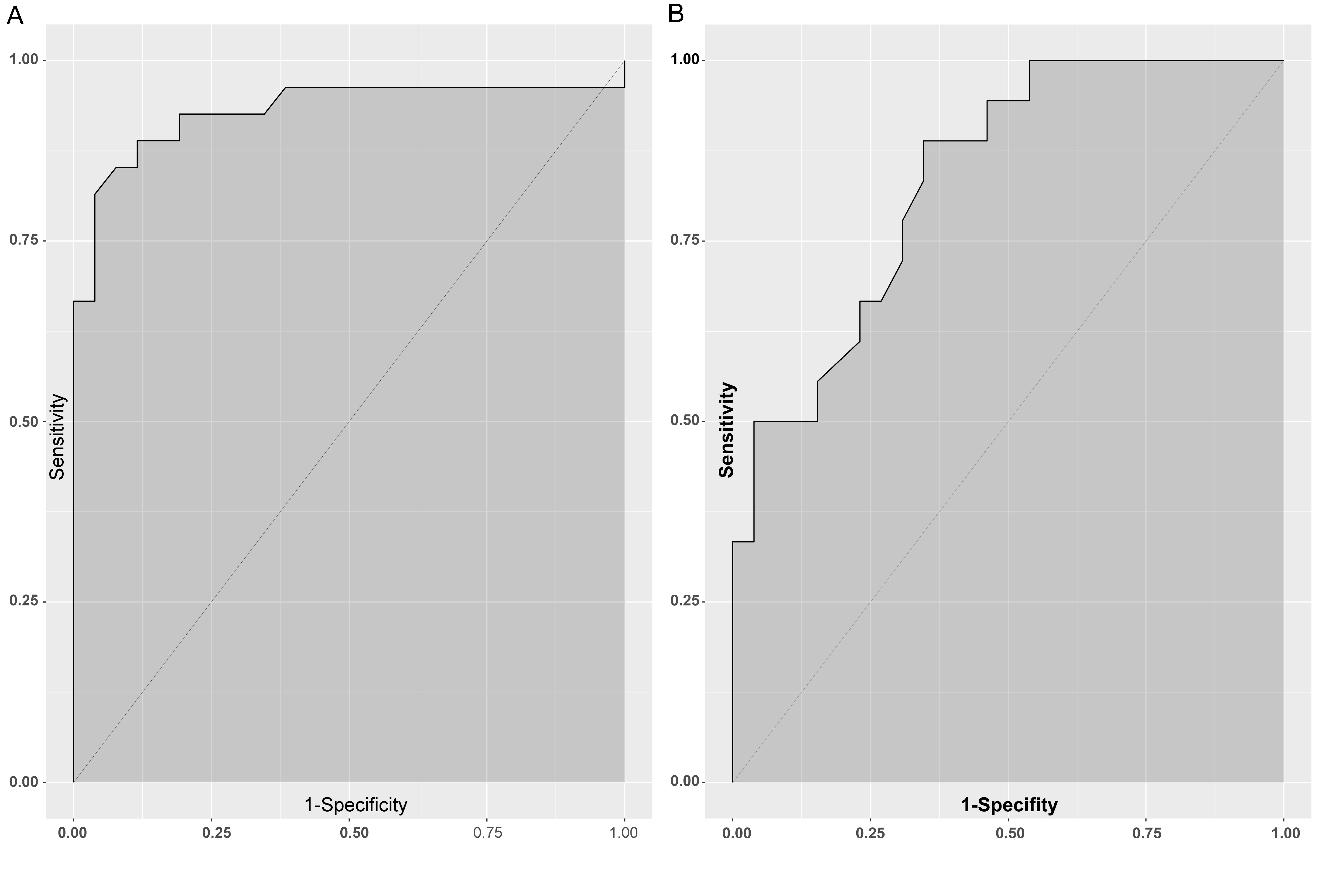
The image quality significantly improved by TSE-DWI (Figure 1 a-d). The signal-to-noise ratio (SNR) for TSE-DWI was lower than SS-EPI but the contrast-to-noise ratio (CNR) was significant higher than SS-EPI (P < 0.001). but the contrast-to-noise ratio (CNR) was significant higher than SS-EPI(P = 0.027). The mean apparent diffusion coefficient (ADC) value on TSE-DWI of the rNPC and post-chemoradiation fibrosis were 0.71×10⁻³ mm2/s and 1.45×10⁻³ mm2/s, respectively, with a significant difference between them (P < 0.001). In comparison, the ADC obtained using SS-EPI are 0.98×10⁻³ mm2/s and 1.17×10⁻³ mm2/s, respectively(P < 0.05). The area under the curve (AUC) for ADC was 0.932 on TSE-DWI and significantly larger than 0.835 on SS-EPI DWI (Figure 2).Discussion
our results demonstrated that the ADC obtained both TSE and SS-EPI showed statistically significant differences between rNPC and post-chemoradiation fibrosis, suggesting that DWI is a useful tool for tissue characterisation and prognostic evaluation in the treatment of NPC. The results were in general agreement with previous studies that used EPI-based DWI to differentiate benign and malignant lesion in head and neck[refs]. Despite the presence of statistically significant differences in overall ADC between the lesions in the present series, the ADC of rNPC on SS-EPI showed a large variation, and partially overlapped with that of post-chemoradiation fibrosis. The large variation are likely due to the measurement errors resulted from magnetic field heterogeneities at the air–bone and air–soft tissue interfaces around the skull base.Conclusion
Our results indicate that TSE-DW imaging of the nasopharynx is preferable and provides more accurate data with higher image quality, along with a significantly improved diagnostic accuracy from 83% to 93% using a clinical high-field-strength (3.0T) MR scanner in the differential diagnosis of rNPC and post-chemoradiation fibrosis.Acknowledgements
This study was supported by the National Scientific Foundation of China (81571664) and the Science and Technology Planning Project of Guangdong Province (2014A020212244, 2016A020216020).References
Ng SH, Chan SC, Yen TC, et al. Comprehensive imaging of residual/ recurrent nasopharyngeal carcinoma using whole-body MRI at 3 T compared with FDG-PET-CT. Eur Radiol 2010; 20:2229-2240.Figures