1805
Preliminary analysis of micro-structural changes in different locations of brain tissue affected by acute ischemic stroke using diffusional kurtosis imagingLiuhong Zhu1, Zhongping Zhang2, Qihua Cheng1, Funan Wang1, and Gang Guo1
1Radiology, Xiamen No.2 Hospital, Xiamen, People's Republic of China, 2MR Research China, GE Healthcare, People's Republic of China
Synopsis
The performance of diffusion kurtosis imaging (DKI) in the analysis of micro-structural changes of brain tissue affected by acute ischemic stroke was explored. 199 lesions in common affected locations were divided into six groups. The value of DKI-derived indices and their changed percentage relative to normal contralateral ROI were calculated. Multiple comparisons among groups indicated that kurtosis indices (especially MK and Ka) showed better performance compared to diffusion indices (ADC, MD, Da and Dr) in detecting structure changes of brain tissues affected by acute ischemic stroke.
Introduction
Diffusional kurtosis imaging (DKI) has been widely used to probe non-Gaussian properties of water diffusion in biological tissues[1-2] and achieved many promising results recently [3-6]. This study aimed to assess the value of DKI in the analysis of micro-structural changes of brain tissue affected by acute ischemic stroke.Methods
One hundred and fifty-six patients (average age: 63.15±12.34 years old, 61 female, 95 male) with acute ischemic stroke underwent routine anatomical MRI and DKI scans (b=0, 1000, 2000s/mm2, 15 directions, TR/TE: 6000/min ms; FOV: 24 cm × 24 cm; thickness/spacing: 5 mm/1.5 mm; number of excitations: 2; matrix: 96×130; the scan time was 6 min and 18 s) from February 2015 to June 2016. Total 199 lesions in common affected locations (Periventricular white matter area (PWM): 52 lesions; corpuscallosum area (CC): 14 lesions; cerebellum area (CB): 29 lesions; basal ganglia area (BG): 21 lesions; brain stem area (BS): 21 lesions; lobes mixed with grey and white matter (LGW): 62 lesions) were outlined. Normal contra-lateral region of interest (ROI) in the opposite mirror region of each lesion was also drawn. We calculated the apparent diffusion coefficient (ADC) and DKI-derived indices including fraction anisotropy (FA), mean diffusion coefficient (MD), axial diffusion coefficient (Da), radial diffusion coeffcient (Dr), mean kurtosis (MK), axial kurtosis (Ka) and radial kurtosis (Kr). The percentage changes of all metrics (ΔFA, ΔMD, ΔDa, ΔDr, ΔMK, ΔKa, ΔKr) with reference to normal contra-lateral ROI were computed. The mean values of these metrics were compared between the two groups using SPSS software and P<0.05 were considered statistical significance).Results
As compared to normal contra-lateral region, ADC, FA, MD, Da and Dr in stroke lesions decreased, MK, Ka and Kr increased in all groups. Diffusional kurtosis exhibited larger fluctuation among groups than diffusivity metrics. Table 1 showed that there was no significant difference of ΔADC among almost all groups (except PWM vs. CB; LGW vs. CB), while there was significant difference of ΔMK among almost all groups (except BG vs. BS; BG vs. LGW; BS vs. LGW) (p<0.05), and ΔKa had the similar results with ΔMK. There was no significant difference of ΔMD and ΔDa among almost all groups (except PWM vs. CB; PWM vs. BG) (p>0.05). ΔDr illustrated no statistical significance among all groups (p>0.05). ΔFA was significantly lower in PWM group and CC group than BG group (p=0.002 and 0.009 respectively). Fig.1 illustrated that the percentage change of diffusional kurtosis in descending order was as following: CC > PWM > BS > LGW > BG > CB.Discussion
The descending order of change percentage of kurtosis-derived parameters (MK and Ka) in different locations illustrated that when the acute ischemic stroke affects tissue mostly contains white matter, the micro-structure changes of tissue are much more complex than other affected locations, which indicated that the order have a trend that the higher proportion of white matter in the tissue, the bigger kurtosis-derived parameters are. What leads to these differences? As we all known, tissues locating at corpuscallosum, brain stem and periventricular white matter areas mainly contained bunches of white matter, while less white matter was found in lobes mixed with grey and white matter. Basal ganglia area contained only grey matter nucleus. The cerebellum consists of a tightly folded layer of cortex with white matter underneath. Combined with the structural characteristics of these tissues and the performances of kurtosis-derived parameters, the results illustrated that there was a trend that when the acute ischemic stroke affects tissue mostly contained white matter, the complexity of micro-structure changes of the tissue was much higher than other affected locations except cerebellum. What have to be motioned is that although we have not distinct evidence on why affected tissue in cerebellum performed quite different with other groups, it must have relationship with its microstructure.Conclusion
When the acute ischemic stroke lesions contain white matter, the complexity of micro-structure changes of the tissue is much higher than other affected locations. DKI could reveal the difference of micro-structure changes among various locations affected by acute ischemic stroke, and diffusional kurtosis perform better than diffusivity among groups.Acknowledgements
No acknowledgement found.References
[1]. Basser, P.J., Jones, D.K. Diffusion-tensor MRI: theory, experimental design and data analysis — a technical review. NMR Biomed. 2002; 15 (7–8), 456–467. [2]. Tuch DS1, Reese TG, Wiegell MR, et al.. Diffusion MRI of complex neural architecture. Neuron. 2003; 40(5), 885–895. [3]. Fieremans E, Jensen J, Helpern JA. White matter characterization with diffusional kurtosis imaging. Neuroimage. 2011; 58(1):177-188. [4]. Yuan J, Yeung DK, Mok GS, Bhatia KS, Wang YX, Ahuja AT, King AD. Non-Guassian analysis of diffusion weighted imaging in head and neck at 3T: A pilot study in patients with nasopharyngeal carcinoma. PLOS ONE. 2014; 8(1):e87024. [5]. Jerry S. Cheung, Enfeng Wang, Eng H Lo, Phillip Zhe Sun. Stratification of heterogeneous diffusion MRI ischemic lesion with kurtosis imaging – Evaluation of mean diffusion and kurtosis MRI mismatch in an animal model of transient focal ischemia. Stroke. 2012; 43(8): 2252–2254. [6]. Umesh Rudrapatna S, Wieloch T, Beirup K, et al.. Can diffusion kurtosis imaging improve the sensitivity and specificity of detecting microstructural alterations in brain tissue chronically after experimental stroke? Comparisons with diffusion tensor imaging and histology. Neuroimage. 2014; 97:363-73.Figures
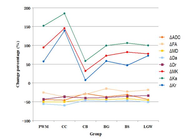
The comparison of ADC and DKI matrices among groups
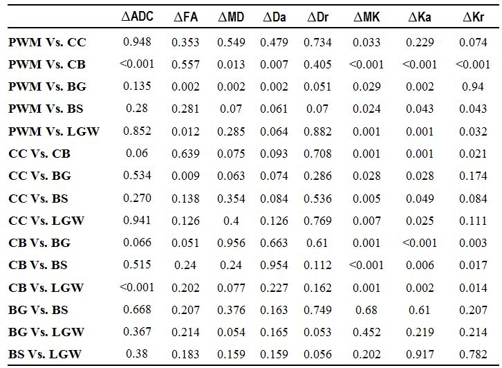
Table 1. Kolmogorov-Smirnov test of
metrics among groups