1795
New approach to improve the reliability of IVIM parameters using computed DWI based on stretched exponential model1Philips Healthcare, Seoul, Korea, Republic of, 2Hanyang University, Seoul, Korea, Republic of, 3Samsung Medical Center, Seoul, Korea, Republic of, 4Philips Healthcare, Tokyo, Japan
Synopsis
We propose a new approach to IVIM analysis using computed DWI (cDWI) based on stretched exponential model. IVIM analysis is widely studied clinically to evaluate tissue perfusion and diffusivity using a range of low and high b values. However, signal attenuation curves of different b values are heterogeneous because of biological effects from multiple components and patient’s motion during the acquisition. So we generated cDWI first using stretched exponential model which can better fit for signal attenuation curve and analyse IVIM parametric maps using cDWI. The proposed approach can fit more robustly the IVIM model parameters.
Introduction
Recently, Intravoxel incoherent motion (IVIM) parameters have been widely used as quantitative imaging biomarkers. But reliable estimation of IVIM parameters is difficult because of the non-linearity of the model, the limited number of low b-values and the low SNR in high b-values [1,2,3]. In this study, we proposed a new approach to estimate IVIM parameters using computed DWI (cDWI) based on the stretched exponential model, having no assumption of the number of intravoxel components. From the cDWI, IVIM parameters could be reliably obtained by increasing the number of b-values and SNR of DWI in low and high b-value.Methods
Acquisition of DWI
Five healthy subjects underwent 3.0T magnetic resonance imaging (Ingenia CX, Philips Healthcare, The Best, Netherlands) using an anterior-posterior coil for single shot-EPI DWI sequence with 10 b-values. (b values = 0, 25, 50, 75, 100, 200, 300, 500, 800, and 1000 s/mm2, TR/TE = 2700ms/67ms, slice thickness = 7mm, matrix size = 128x126, FOV = 40x40cm2, the number of slices = 29, and total scan time = 2.5 minutes).
MRI data analysis
cDWI using Stretched exponential model
For cDW images, the stretched-exponential model [4] was used.
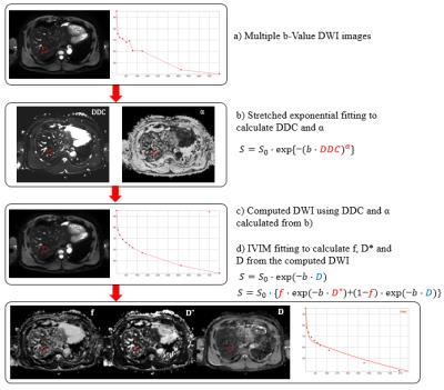
$$$\frac{S(b)}{S_{0}}=\exp(-{(bDDC)^\alpha)}$$$, where $$$\alpha$$$ is the stretching parameter, which characterizes the deviation of the signal attenuation from the mono-exponential model. A value of $$$\alpha$$$ that is near one indicates high homogeneity in apparent diffusion, whereas a low value of $$$\alpha$$$ result from non-exponential model caused by the addition of multiple components. The DDC is the distributed diffusion coefficient, which is similar to D parameter in mono-exponential model. For each voxel, the value of $$$\frac{S(b)}{S_{0}}$$$ was computed using estimated DDC and $$$\alpha$$$ values, and then the cDWI data (S(b)) were estimated.
IVIM parameters
For estimating IVIM parameters, the bi-exponential model was used.
$$$\frac{S(b)}{S_{0}}=f\times\exp(-bD*)+(1-f)\times\exp(-bD)$$$
, where f is the perfusion fraction, D is the diffusion coefficient and D* is the pseudo-diffusion coefficient. To obtain the IVIM parameters, the IVIM signal equation above was calculated by means of the Luciani simplified method [2,5] using home-built SW (Diffusion analysis software, EXPRESS 2.0, Philips Healthcare, Korea). To estimate D parameters, first, the curve was fitted for b>200 s/mm2 (D* is significantly greater than D). And then the curve was fitted for f and D* over all values of b, while keeping D constant. This simplified two-step method was used to increase robustness under biological conditions. For comparison, we compared the IVIM parameters estimated from raw DWI and the cDWI calculated by the stretched exponential model.
Results
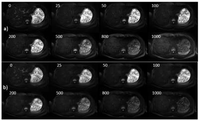
Fig. 1 showed the diagram for our proposed method. The raw DWI data was fitted based on the stretched exponential model and cDWI data were estimated. In low and high b-values, we found the low SNR and the regional heterogeneity in raw DWI data, but these noisy patterns were reduced in the cDWI data based on the stretched exponential model as shown in Fig 2. The cDWI data were well fitted on IVIM model rather than the raw DWI data, showing less noisy patterns on the IVIM parametric maps, especially in D* map (Fig. 3). For quantitative analysis, we measured the IVIM parameters from 75 ROIs across 5 subjects. From the results, we found that 7.3% difference for f (293.7±121.58 for raw DWI, and 306.2±111.6 for cDWI), 8.0% difference for D (771.8±250 for raw DWI, and 692.7±260.8 for cDWI), and 47.4% difference for D* (1100.3±4159.359 for raw DWI, and 199.7±851.3 for cDWI) (x $$$10^{-3} mm^2/s$$$). Fig. 4 showed that the value of IVIM parameters estimated from cDWI is more uniform distribution than those estimated from raw DWI.Discussion and conclusion
In this preliminary study, we proposed a new method for reliable estimation of IVIM parameters using cDWI based on the stretched exponential model. Our findings showed the low variance of IVIM parameters, especially in the D* parameter, showing the reliable estimates of IVIM parameters on liver. The unreliable D* parameter estimated from the raw DWI data may be due to the low diffusion effect using small diffusion gradient with more sensitive to biological conditions and patient motion in low b-value. Using cDWI data, we believed that the low SNR over all b values and various artifacts in low b value could be compensated, leading more reliable estimates of IVIM parameters. In this study, we tested the feasibility of our method in liver parenchyma for health subjects, and further study will be needed to validate our method in patients with various liver diseases.Acknowledgements
No acknowledgement found.References
1. Le Bihan D, MR imaging of intravoxel incoherent motions: application to diffusion and perfusion in neurologic disorders.Radiology. 1986 Nov; 161(2):401-7.
2. Luciani A, Vignaud A, Cavet M, et al. Liver cirrhosis: intravoxel incoherent motion MR imaging pilot study. Radiology 2008;249(3):891– 899.
3.Dow-Mu Koh, David J. Collins, Matthew R. Orton et al. Intravoxel Incoherent Motion in Body Diffusion-Weighted MRI: Reality and Challenges. AJR:196, June 2011
4.Bennett KM, Schmainda KM, Bennett RT et al. Characterization of continuously distributed cortical water diffusion rates with a stretched-exponential model. Magn Reson Med. 2003 Oct;50(4):727-34.
5. Seber GA, Wild CJ. Nonlinear regression. Hoboken, NJ: Wiley-Interscience; 2003.
6. Sigmund et al. Intravoxel incoherent motion and diffusion-tensor imaging in renal tissue under hydration and furosemide flow challenges. Radiology. 2012 Jun;263(3):758-69
Figures


